Bioluminescence Vs Fluorescence Imaging
The ability to visualize biological processes in living organisms has revolutionized medical research and diagnostics. Two powerful imaging techniques, bioluminescence and fluorescence imaging, are at the forefront of this revolution, offering unprecedented insights into the inner workings of cells, tissues, and entire organisms. However, these methods, while sharing the common goal of visualization, operate on fundamentally different principles and offer distinct advantages and limitations.
Understanding the nuances between bioluminescence and fluorescence is crucial for researchers selecting the appropriate imaging modality for their specific experimental needs. This article will delve into the core mechanisms of each technique, compare their applications in diverse fields such as drug discovery and cancer research, and explore emerging trends that promise to further enhance their capabilities. The ultimate goal is to provide a comprehensive overview of these vital tools for biomedical exploration.
Understanding the Fundamentals
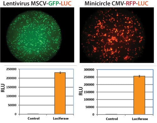
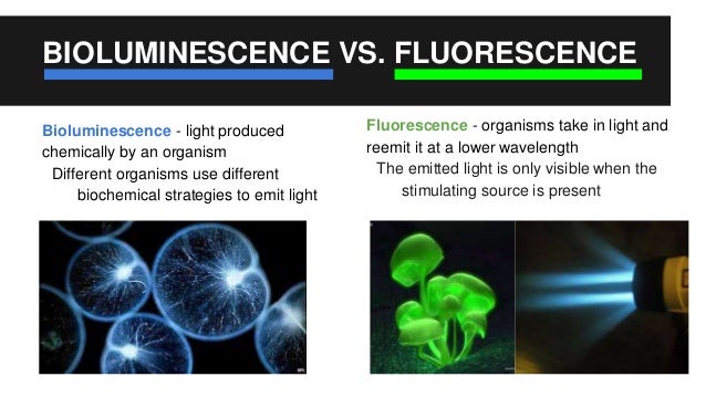
Bioluminescence imaging (BLI) relies on the inherent ability of certain living organisms, such as fireflies, to produce light through biochemical reactions. This process typically involves the enzyme luciferase, which catalyzes a reaction with a substrate like luciferin, resulting in the emission of photons. The light emitted is then captured by highly sensitive cameras.
Unlike BLI, fluorescence imaging (FLI) requires an external light source to excite fluorescent molecules, known as fluorophores. These fluorophores absorb light at a specific wavelength and then emit light at a longer wavelength, which is detected by imaging systems. The difference in wavelengths allows for selective detection of the emitted light, separating it from the excitation source.
Advantages and Disadvantages
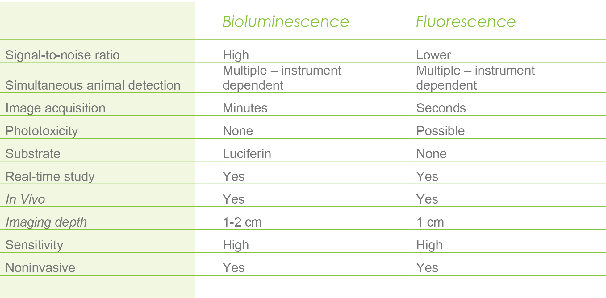
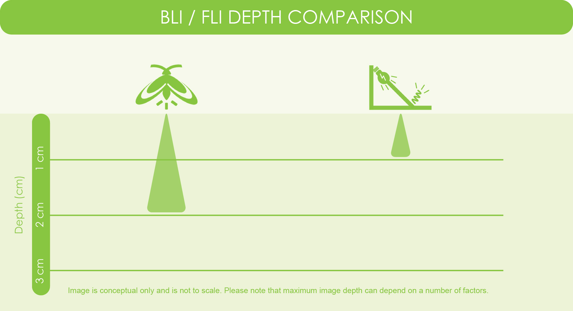
BLI offers several advantages, including high sensitivity and low background signal. Because the light is generated within the organism, there is minimal interference from external light sources, leading to clearer images, according to a report published in the journal Nature Methods. This is particularly beneficial for deep tissue imaging.
However, BLI also has limitations, such as the requirement for genetic modification to introduce the luciferase gene into the organism or cells of interest. Furthermore, the emitted light is typically weaker than that of fluorescence, and the color range is more limited. In vivo studies have shown BLI signal decreases with depth.
FLI boasts a wider range of available fluorophores, enabling researchers to target specific cellular components or processes with greater precision. Multiple fluorophores with different excitation and emission spectra can be used simultaneously for multiplexed imaging. This allows researchers to monitor multiple parameters at the same time, in vivo.
The main drawback of FLI is its susceptibility to autofluorescence, where naturally occurring molecules in the tissue emit light upon excitation, creating background noise. This can reduce the signal-to-noise ratio and make it difficult to visualize faint signals, according to the National Institutes of Health.
Applications in Research and Medicine
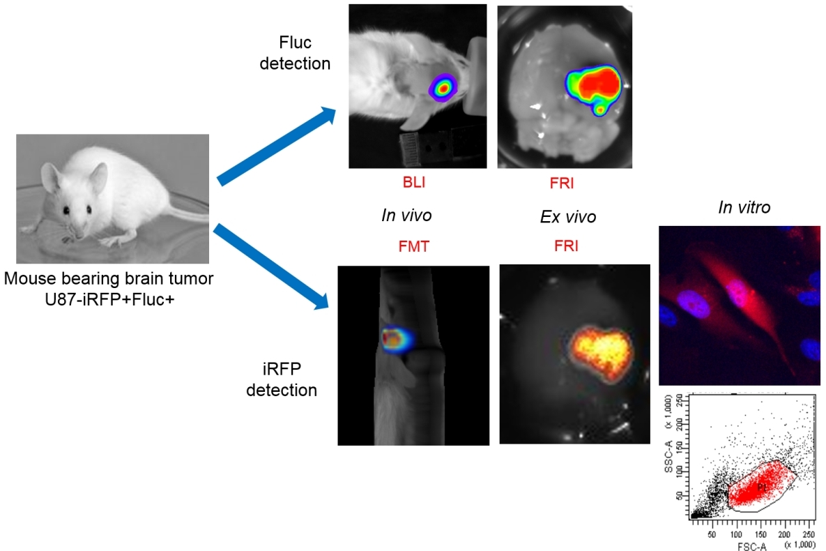
Both BLI and FLI are widely used in preclinical research, particularly in drug discovery and development. BLI is commonly used to monitor tumor growth and metastasis in animal models, assess the efficacy of cancer therapies, and track gene expression. Studies published in Cancer Research show the importance of non-invasive tumor monitoring.
FLI is frequently employed to study cellular signaling pathways, protein-protein interactions, and enzyme activity. It plays a crucial role in understanding fundamental biological processes and identifying potential drug targets. Studies in Cell shows the use of fluorescence to track complex molecular processes.
In clinical settings, FLI is gaining traction for image-guided surgery and diagnostics. Fluorescent dyes can be used to highlight tumors or other abnormal tissues during surgical procedures, improving precision and reducing the risk of complications. According to a study published in The Lancet, it can assist in the detection of cancerous tissues.
Emerging Trends and Future Directions
Researchers are constantly developing new and improved bioluminescent and fluorescent probes with enhanced brightness, stability, and specificity. Genetically encoded fluorescent indicators (GEFIs) are becoming increasingly popular for long-term monitoring of cellular activity in vivo.
Advanced imaging techniques, such as two-photon microscopy and bioluminescence resonance energy transfer (BRET), are expanding the capabilities of BLI and FLI. These methods offer improved spatial resolution, penetration depth, and sensitivity. BRET is a method which involves the transfer of energy from a bioluminescent donor to a fluorescent acceptor.
Furthermore, the integration of artificial intelligence and machine learning algorithms is transforming the analysis of bioluminescent and fluorescent images. AI-powered tools can automate image processing, identify subtle patterns, and extract quantitative data with greater accuracy. These methods can make the imaging process more efficient.
Conclusion
Bioluminescence and fluorescence imaging are indispensable tools for biological research and medical diagnostics. While each technique has its own strengths and weaknesses, both offer invaluable insights into the complexities of living systems. As technology continues to advance, these imaging modalities will undoubtedly play an even greater role in unraveling the mysteries of biology and improving human health.


.jpg)

.jpg)