Cell Bodies Of Sensory Neurons Are Located In The
Imagine walking barefoot on a warm beach, the fine sand yielding under your feet. The gentle caress of a cool breeze whispers past, and the distant cry of seagulls fills the air. Every sensation, from the pressure on your soles to the scent of salt in the air, is a message relayed by a complex network within you, a silent symphony of communication happening every moment.
At the heart of this sensory marvel lies a fundamental question: where are the control centers, the headquarters if you will, for the neurons responsible for these sensations? The answer is elegantly simple: the cell bodies of sensory neurons reside predominantly in structures called ganglia, specifically the dorsal root ganglia for many of our bodily sensations and cranial nerve ganglia for sensations from the head and face. Understanding this seemingly small detail unlocks a deeper appreciation of how our nervous system orchestrates our experience of the world.
The Sensory Superhighway: A Journey From Sensation to Perception
To truly grasp the significance of the dorsal root ganglia (DRG) and cranial nerve ganglia, let's embark on a journey tracing the path of a sensory signal. It all starts at the sensory receptors, specialized cells that detect stimuli – whether it's the touch of a feather, the heat of a flame, or the sound of music.
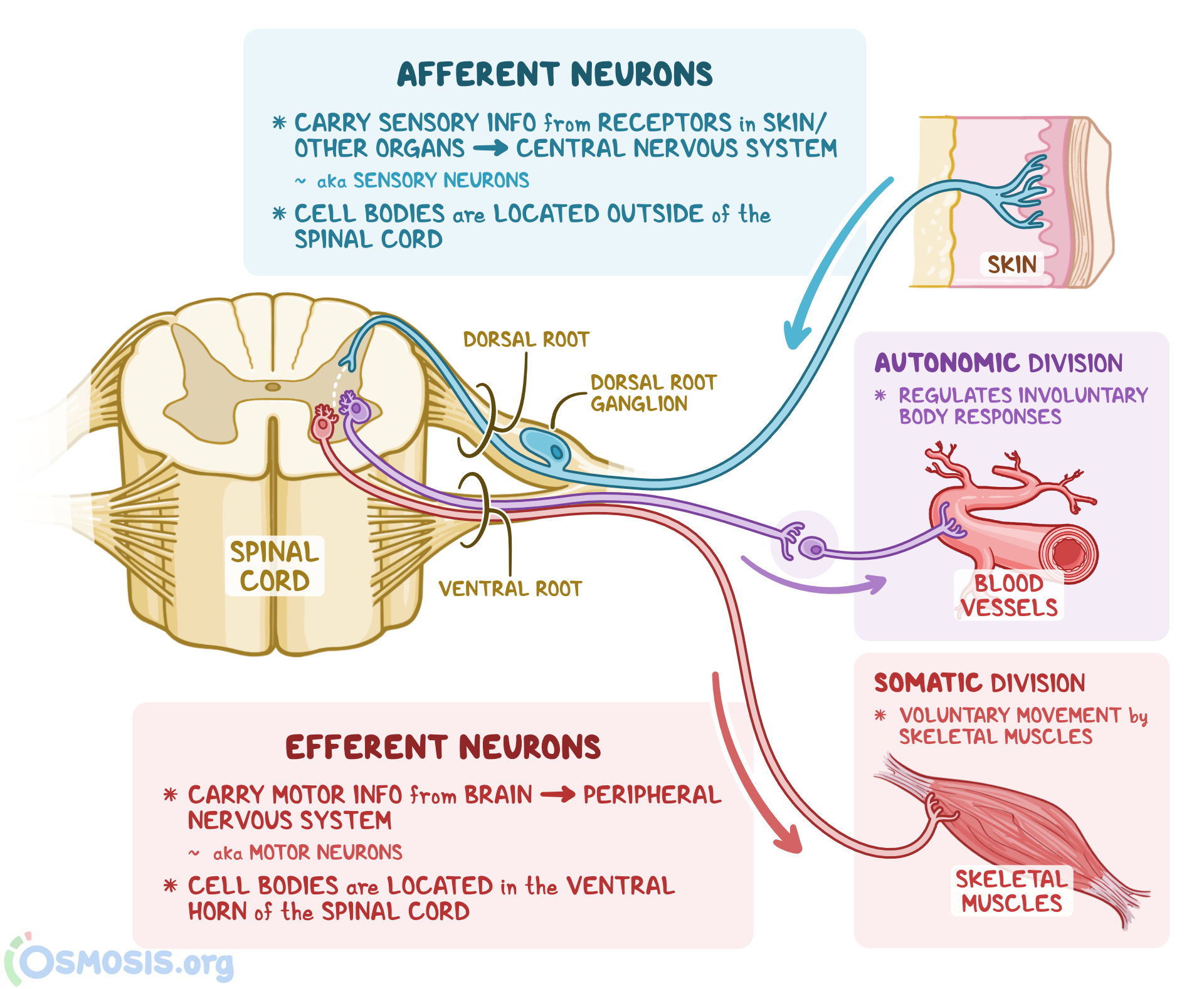
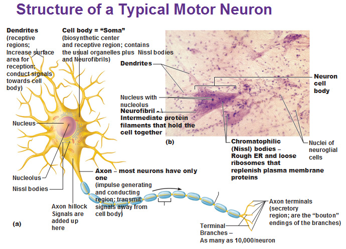
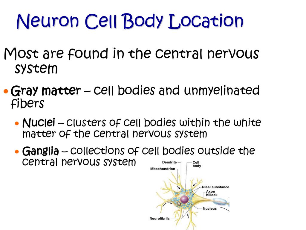
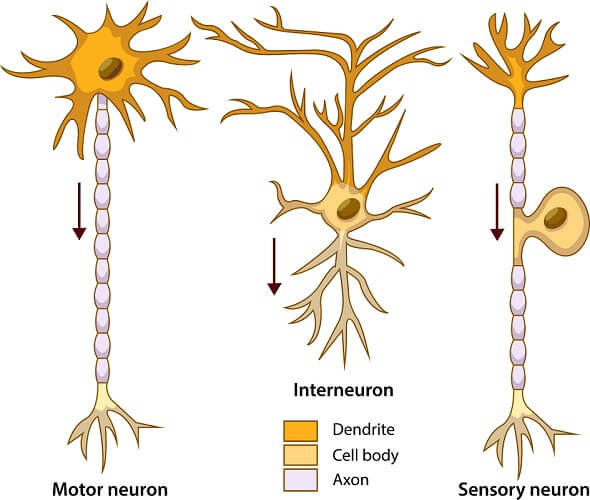
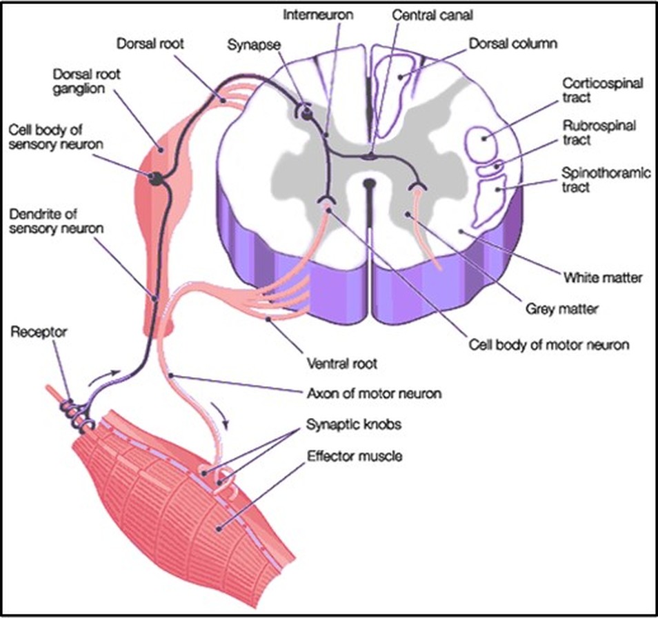
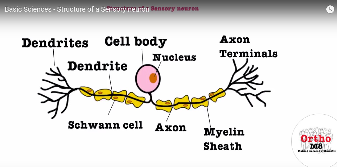
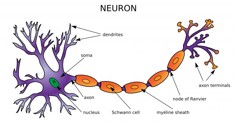
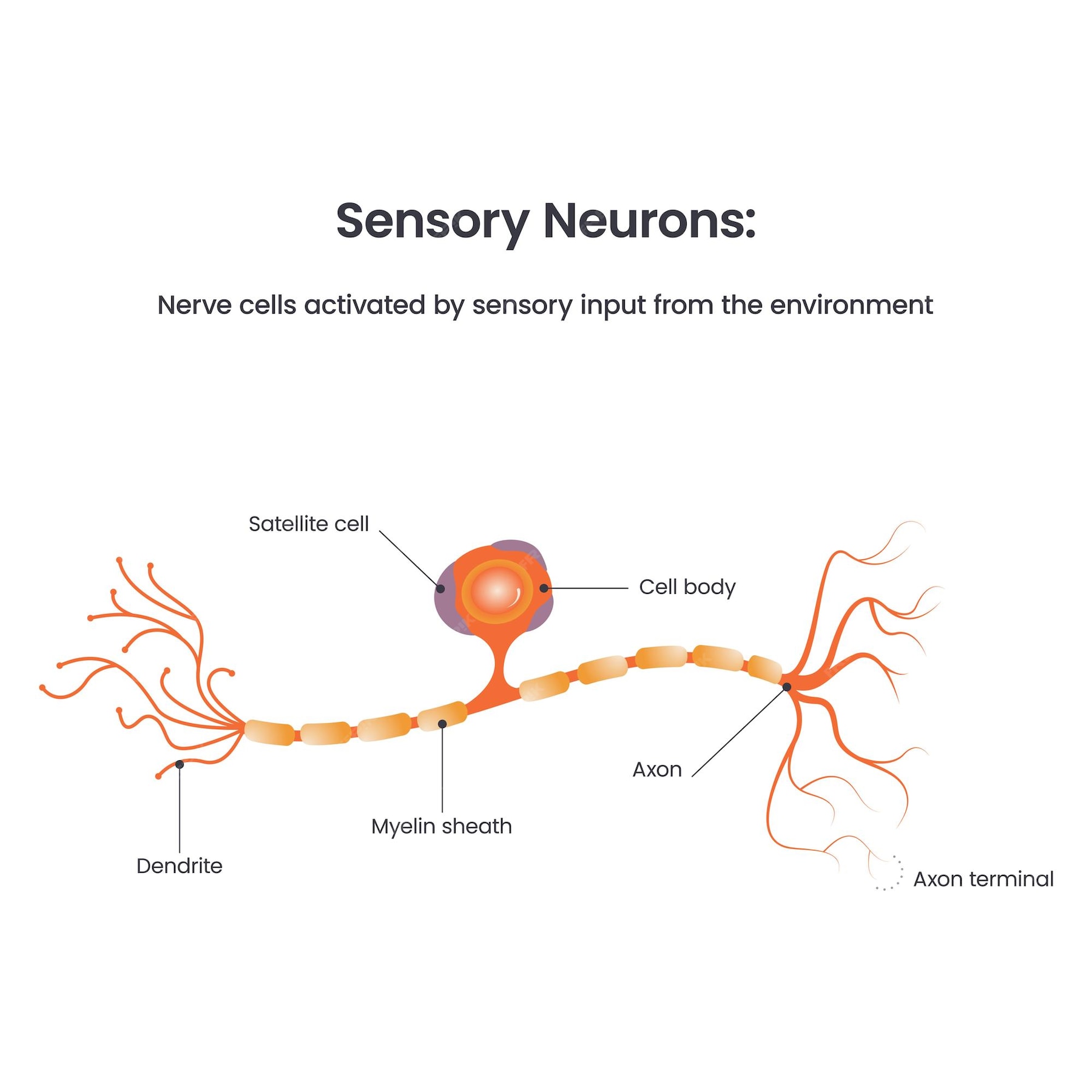
These receptors transform the physical stimulus into an electrical signal, which then travels along the sensory neuron. The sensory neuron, unlike typical neurons, has a unique structure. Its cell body, containing the nucleus and essential cellular machinery, is not located within the central nervous system (CNS) but resides outside, nestled within the protective embrace of a ganglion.
Dorsal Root Ganglia: Guardians of Bodily Sensations
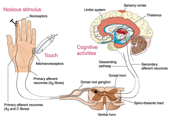
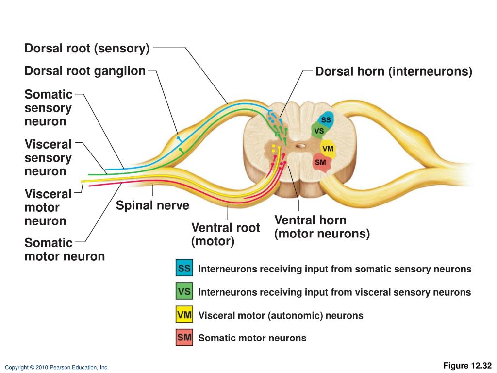
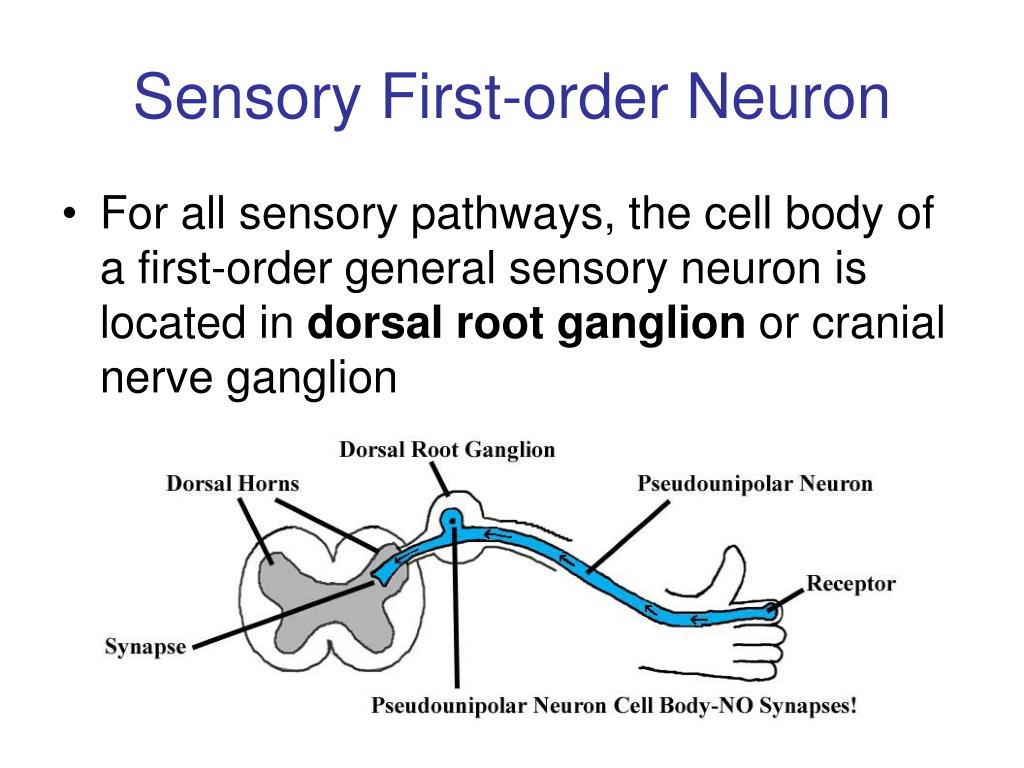
The dorsal root ganglia are clusters of sensory neuron cell bodies located along the dorsal roots of the spinal cord. These roots are the pathways through which sensory information enters the spinal cord from the body.
Think of the spinal cord as the information superhighway leading to the brain. The dorsal roots are the on-ramps, and the dorsal root ganglia are like service stations along the way, providing essential support and maintenance for the sensory neurons.
Each dorsal root ganglion contains the cell bodies of sensory neurons that carry information about touch, temperature, pain, and proprioception (the sense of body position). These neurons gather signals from all over the body, excluding the head and some parts of the face.
Cranial Nerve Ganglia: Sentinels of the Head and Face
While the dorsal root ganglia handle the bulk of bodily sensations, specialized ganglia associated with the cranial nerves take responsibility for the head and face. These cranial nerve ganglia serve a similar function, housing the cell bodies of sensory neurons that innervate the face, mouth, eyes, and other structures in the head.
For instance, the trigeminal ganglion, the largest of the cranial nerve ganglia, contains the cell bodies of sensory neurons responsible for touch, pain, and temperature sensations in the face. Other cranial nerve ganglia are associated with taste, smell, and hearing.
"The anatomical location of these cell bodies outside the central nervous system provides a critical layer of protection," explains Dr. Anya Sharma, a neuroscientist specializing in sensory systems. "By keeping the cell bodies away from the immediate vicinity of the spinal cord and brain, they are somewhat shielded from potential damage and inflammation."
Why Does Location Matter? The Significance of Ganglia
The strategic placement of sensory neuron cell bodies in ganglia is more than just an anatomical curiosity; it has important functional implications. One key advantage is protection. The ganglia provide a physical barrier, shielding the cell bodies from injury and the potentially harmful effects of inflammation within the central nervous system.
Furthermore, the location of the cell bodies outside the CNS simplifies the organization and routing of sensory information. It allows for a more efficient and direct connection between the sensory receptors in the periphery and the spinal cord or brainstem.
This arrangement also contributes to the modularity of the nervous system. Each ganglion can be viewed as a distinct processing unit, dedicated to specific types of sensory information from a particular region of the body.
Clinical Relevance: When Ganglia Go Awry
Understanding the anatomy and function of sensory ganglia is crucial for diagnosing and treating various neurological conditions. Damage or dysfunction of these ganglia can lead to a range of sensory deficits, including numbness, tingling, pain, and loss of proprioception.
For example, shingles, a viral infection caused by the varicella-zoster virus (the same virus that causes chickenpox), can affect the dorsal root ganglia, causing intense pain and a characteristic rash along the affected dermatome (the area of skin innervated by a single spinal nerve). Postherpetic neuralgia, a chronic pain condition that can develop after shingles, is thought to be related to persistent damage to the sensory neurons within the dorsal root ganglia.
Similarly, certain autoimmune disorders, such as Guillain-Barré syndrome, can target the peripheral nerves and ganglia, leading to sensory and motor impairments. Advances in neuroimaging techniques, such as MRI, are enabling clinicians to visualize the ganglia and detect abnormalities that may be indicative of underlying neurological conditions.
Looking Ahead: Future Directions in Ganglion Research
Research on sensory ganglia is an active and evolving field. Scientists are exploring the intricate molecular mechanisms that regulate the development, function, and survival of sensory neurons within the ganglia.
One promising area of investigation is the development of novel therapies for chronic pain. Researchers are investigating targeted drug delivery methods to deliver pain-relieving medications directly to the dorsal root ganglia, minimizing systemic side effects.
Another exciting avenue of research is the use of stem cells to regenerate damaged sensory neurons within the ganglia. These efforts hold the potential to restore sensory function in patients with nerve injuries or neurodegenerative diseases.
As we continue to unravel the mysteries of the nervous system, the humble ganglion, often overlooked, emerges as a critical player in our ability to perceive and interact with the world around us. Its strategic location, protective function, and clinical significance underscore its importance in maintaining our sensory well-being.