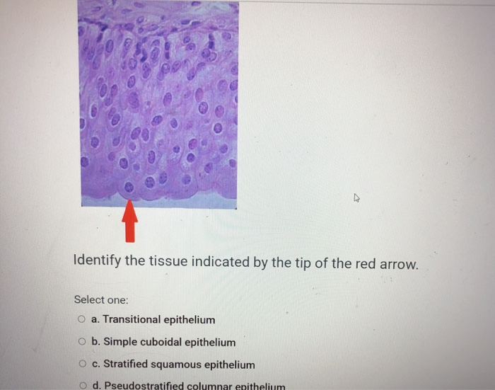
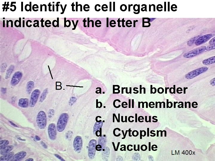
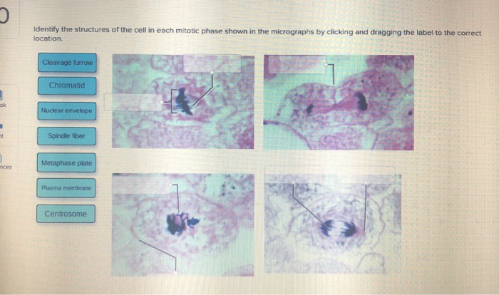
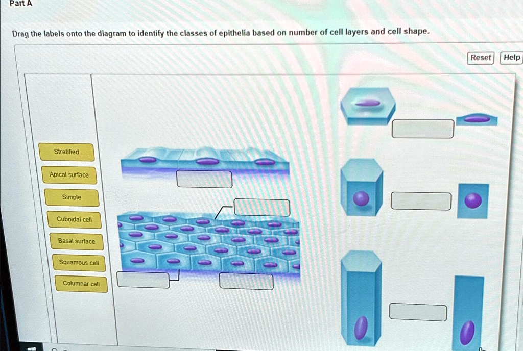
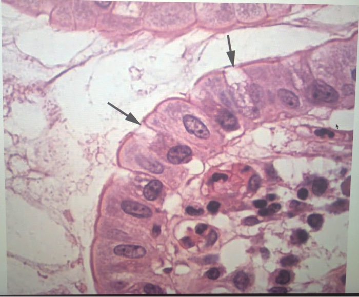
Identify The Shape Of The Cell Indicated By The Line
Panic grips the scientific community as a mysterious anomaly plagues cellular imaging: Lines superimposed on micrographs are defying categorization, triggering widespread diagnostic uncertainty.
This crisis, dubbed "ShapeGate" by some, centers on the inability to accurately identify cell shapes due to these disruptive linear markings. The incident is impacting critical research and diagnostic processes across various fields, demanding immediate resolution.
The Anomaly: Lines of Unknown Origin
The issue first surfaced at the National Institute of Cellular Biology (NICB) on October 26, 2024, during a routine analysis of epithelial cell cultures. Researchers noticed a sharp, unwavering line bisecting multiple cells in the microscopic images.
Initially dismissed as an artifact, the phenomenon quickly spread, appearing in labs at Harvard Medical School and the University of Tokyo. Images from diverse cell types – neurons, erythrocytes, fibroblasts – were all affected. The lines are consistent in appearance: straight, single pixel width, and always present irrespective of staining techniques or imaging modalities.
“We’ve tried everything. Different microscopes, different preparation techniques, different labs,” stated Dr. Anya Sharma, lead researcher at NICB. “Nothing eliminates these lines.”
Impact on Diagnostic Procedures
The most pressing concern is the effect on medical diagnostics. Cell shape is a crucial indicator for identifying various diseases, including cancer and infectious diseases.
“Our ability to distinguish between healthy and cancerous cells based on morphology is compromised,” explains Dr. Kenji Tanaka, head pathologist at Tokyo University Hospital. “The lines obscure the true shape, leading to potential misdiagnosis.”
The World Health Organization (WHO) issued a preliminary advisory urging caution in interpreting cellular images affected by the anomaly. Dr. Maria Rodriguez, spokesperson for the WHO, emphasized the need for rigorous validation of diagnostic results.
The Search for Answers: Technical Glitch or Something More?
The investigation is focusing on two primary hypotheses: a systemic software malfunction and an external environmental factor.
Teams of software engineers are scrutinizing imaging software and analysis tools for potential bugs. Early investigations have ruled out common issues like corrupted files or driver incompatibilities.
The possibility of an external influence, such as electromagnetic interference, is also being explored. Specialized equipment has been deployed at affected labs to monitor environmental conditions.
“We are considering all possibilities, no matter how improbable they may seem,” asserted Dr. David Chen, cybersecurity expert assisting the investigation. He adds, “The coordinated nature of the appearance suggests a non-random cause.”
Global Collaboration Underway
A global task force, coordinated by the International Council of Scientific Unions (ICSU), has been formed to tackle the issue. Researchers from over 40 countries are sharing data and expertise.
The task force is employing advanced image analysis techniques, including artificial intelligence, to try and filter out the lines and reconstruct the original cell shapes. They're also performing blind tests, attempting to accurately classify cells with and without the anomaly.
The European Molecular Biology Laboratory (EMBL) is leading the effort to establish standardized protocols for image acquisition and analysis to minimize variability and identify potential sources of error.
Collaboration is key. We need to pool our resources and knowledge to solve this problem quickly,” stated Professor Isabelle Dubois, head of the EMBL task force.
Immediate Actions and Ongoing Developments
Until the cause is identified, diagnostic labs are advised to use multiple independent methods for cell identification and confirmation. Retrospective analysis of previously acquired images is also recommended.
The ICSU task force has scheduled a press conference for November 15, 2024, to provide an update on the investigation. Further information is expected to be released on their website.
The situation remains critical, with the potential to disrupt vital scientific and medical activities. The global scientific community is united in its determination to resolve this perplexing anomaly and restore confidence in cellular imaging.