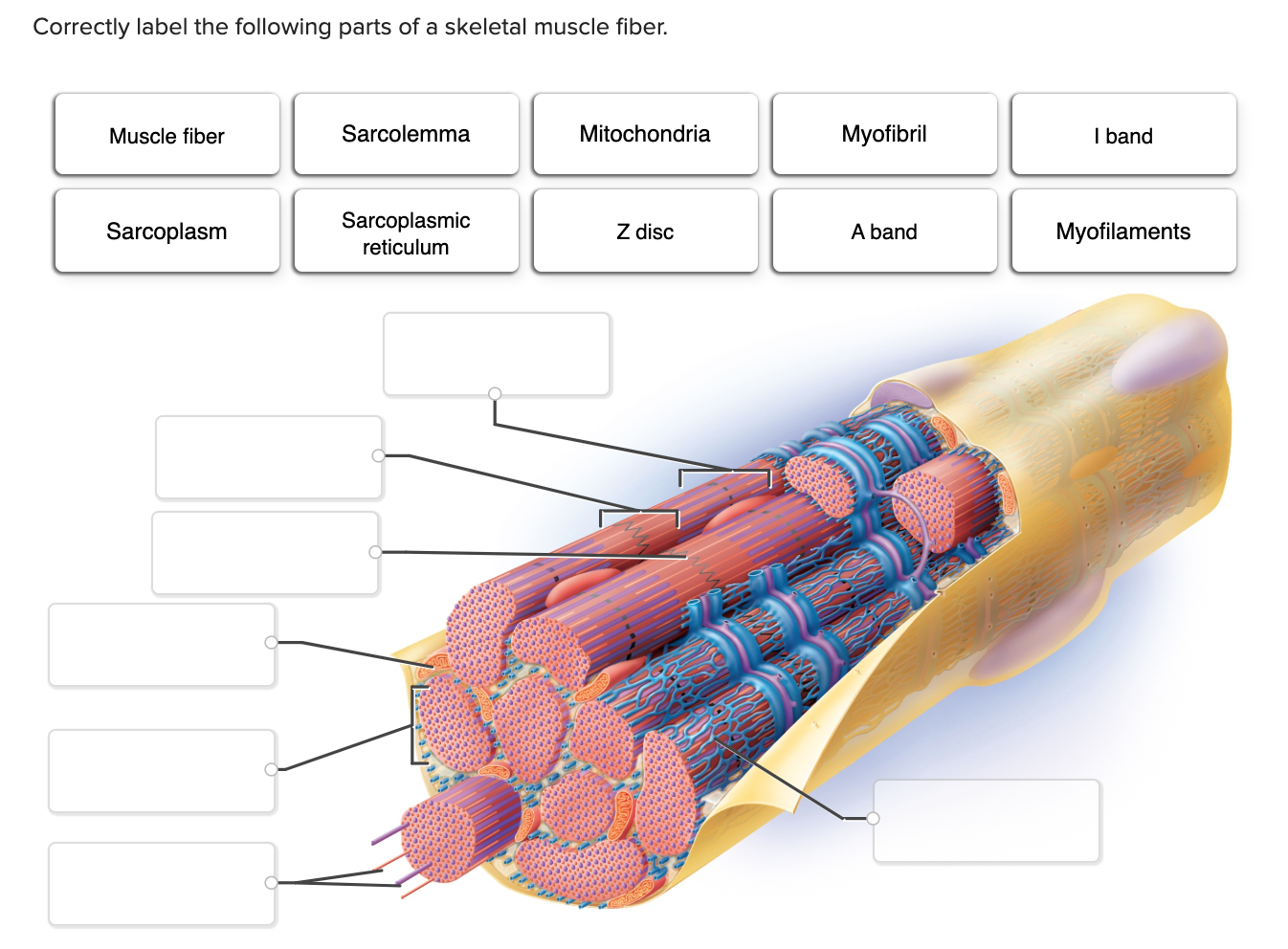
Match The Component Of A Muscle Cell With Its Description.

Imagine peering through a microscope, the intricate world of the human body unfolding before your eyes. Within each twitch, flex, and hold, lies a universe of cellular activity. The smooth, coordinated movements you take for granted are actually the result of a complex symphony of tiny structures working in perfect harmony within your muscle cells.
This article delves into the fascinating landscape of muscle cell components, offering a clear understanding of how each part contributes to the overall function of muscle contraction and movement. We'll explore the key players in this biological process, matching each component with its specific role.
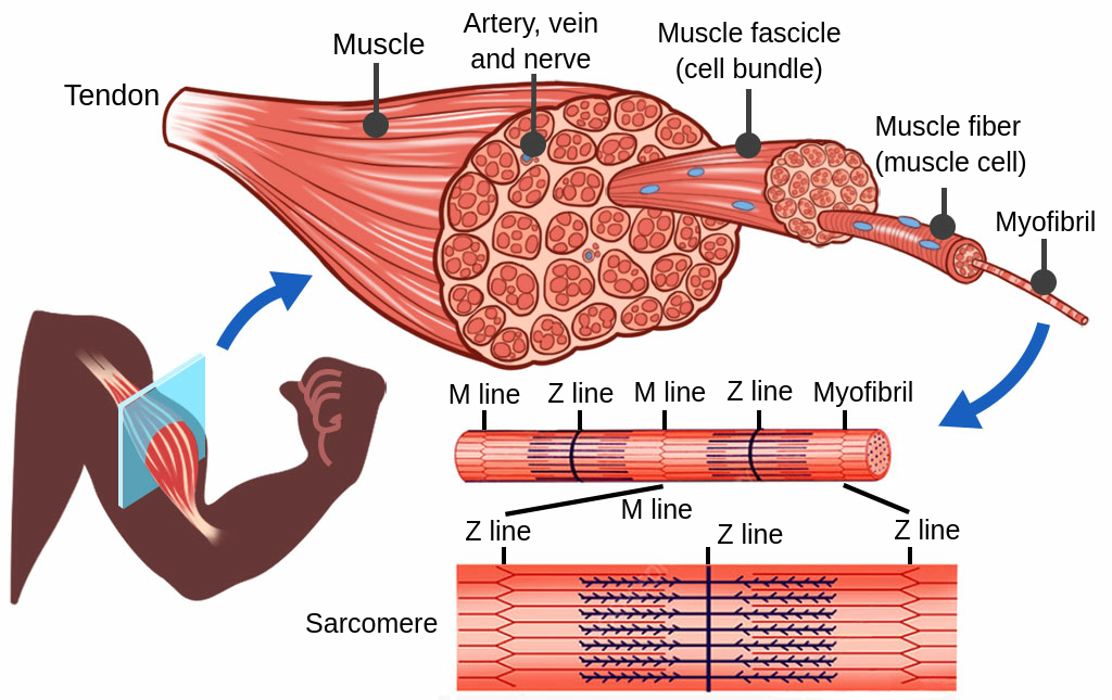
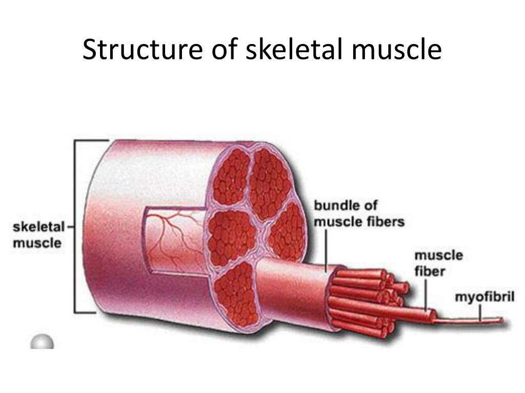
Understanding Muscle Cell Architecture
Muscle cells, also known as muscle fibers, are highly specialized cells responsible for generating force and enabling movement. These elongated cells are packed with proteins and structures that facilitate the sliding of filaments, leading to muscle contraction.
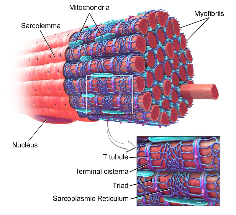
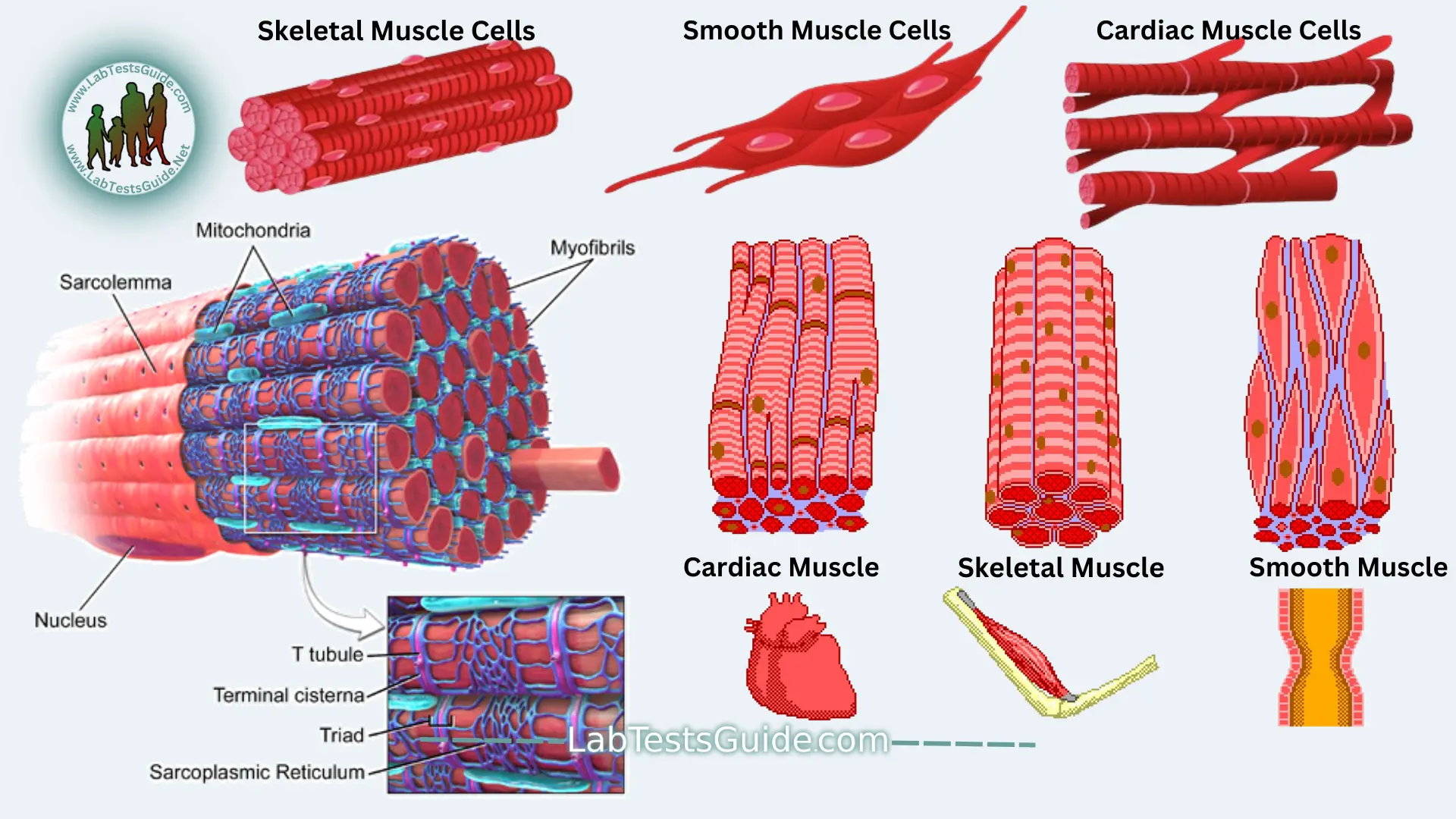
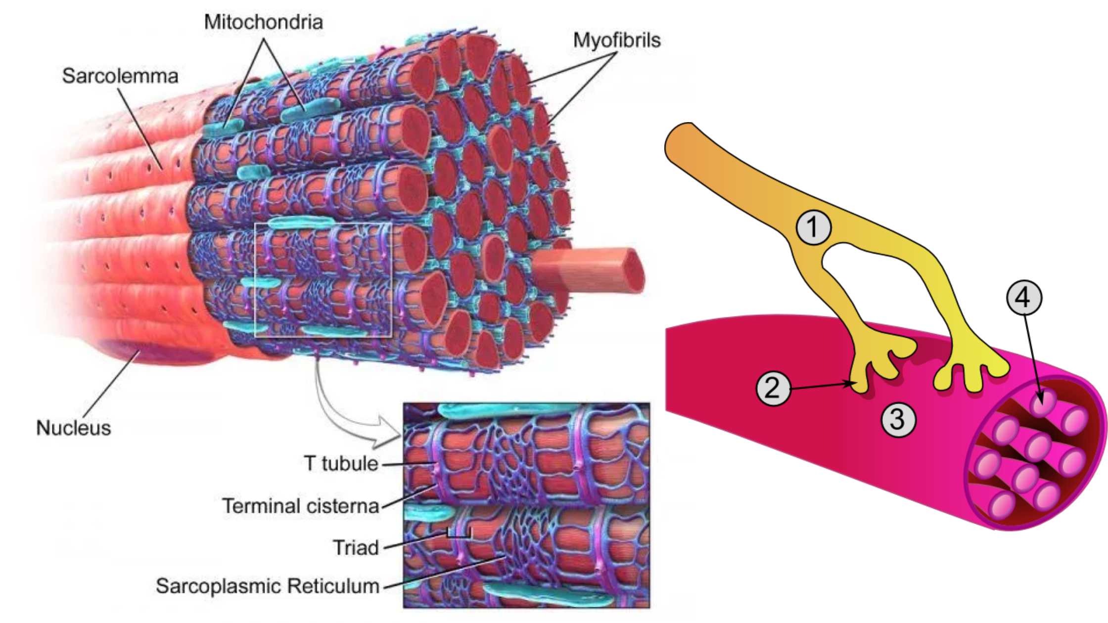
The Sarcolemma: The Cell's Guardian
The sarcolemma is the cell membrane of a muscle fiber, forming a protective barrier that encloses the cytoplasm, known as the sarcoplasm. It's not just a passive barrier; it also plays a crucial role in conducting electrical signals that initiate muscle contraction.
Sarcoplasmic Reticulum: Calcium's Reservoir
Deep within the sarcoplasm lies the sarcoplasmic reticulum (SR), a network of interconnected tubules responsible for storing and releasing calcium ions. Calcium ions are essential for triggering muscle contraction.
T-Tubules: Conducting the Signal
To ensure that every part of the muscle fiber receives the signal to contract, the sarcolemma forms inward extensions called transverse tubules, or T-tubules. These tubules run perpendicular to the muscle fiber and allow the action potential to rapidly spread throughout the cell.
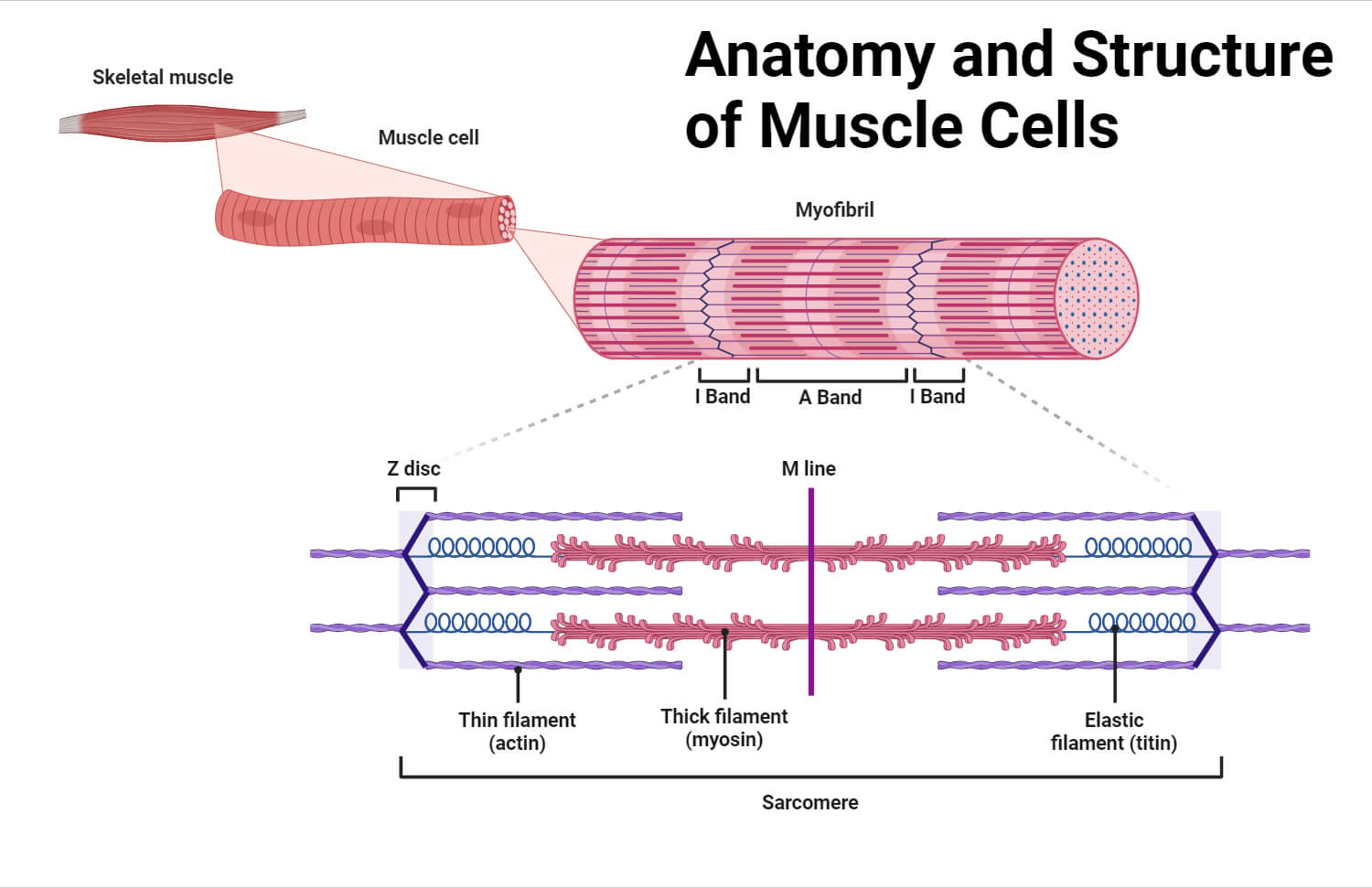
Myofibrils: The Contractile Engine
Muscle fibers are filled with long, cylindrical structures called myofibrils. These are the fundamental contractile units of the muscle cell, and they are responsible for generating the force that produces movement.
Myofibrils are composed of repeating units called sarcomeres, the basic functional unit of muscle contraction. Think of sarcomeres as individual links in a chain, each contributing to the overall strength of the muscle.
Myofilaments: The Proteins of Contraction
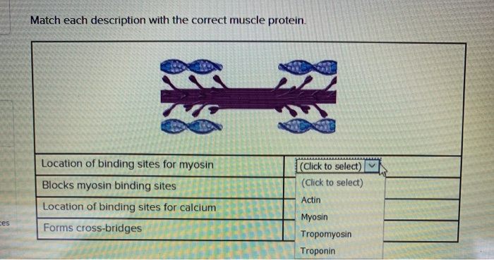
Within each sarcomere, you'll find two primary types of protein filaments: actin (thin filaments) and myosin (thick filaments). These filaments interact to produce muscle contraction through a process called the sliding filament theory.
Actin filaments are composed of globular actin molecules arranged in a helical structure. They also contain proteins called tropomyosin and troponin, which regulate the interaction between actin and myosin.
Myosin filaments are composed of myosin molecules, each with a head that can bind to actin. The myosin heads act as tiny motors, pulling the actin filaments and causing the sarcomere to shorten.
The Sliding Filament Theory: A Step-by-Step Look
The sliding filament theory explains how muscle contraction occurs. Here's a simplified breakdown:
1. Nerve Impulse: A nerve impulse arrives at the neuromuscular junction, triggering the release of acetylcholine.
2. Action Potential: Acetylcholine binds to receptors on the sarcolemma, generating an action potential that travels along the sarcolemma and down the T-tubules.
3. Calcium Release: The action potential triggers the release of calcium ions from the sarcoplasmic reticulum.
4. Binding Site Exposure: Calcium ions bind to troponin, causing a conformational change in tropomyosin, which exposes the binding sites on actin.
5. Cross-Bridge Formation: Myosin heads bind to the exposed binding sites on actin, forming cross-bridges.
6. Power Stroke: The myosin heads pivot, pulling the actin filaments toward the center of the sarcomere, shortening the sarcomere.
7. ATP Binding and Detachment: ATP binds to the myosin heads, causing them to detach from actin.
8. Myosin Reactivation: ATP is hydrolyzed, providing energy to re-cock the myosin heads, ready to bind to actin again.
This cycle repeats as long as calcium ions are present and ATP is available, resulting in continuous muscle contraction. When the nerve impulse stops, calcium ions are pumped back into the sarcoplasmic reticulum, and the muscle relaxes.
Matching Components to Descriptions: A Quiz
Let's test your understanding. Match the muscle cell component with its description:
1. Sarcolemma 2. Sarcoplasmic Reticulum 3. T-Tubules 4. Myofibrils 5. Sarcomere 6. Actin 7. Myosin
A. The basic functional unit of muscle contraction. B. The cell membrane of a muscle fiber. C. Stores and releases calcium ions. D. Contains thin filaments. E. Contains thick filaments. F. Conducts action potentials throughout the muscle fiber. G. The contractile units of the muscle cell.
Answers: 1-B, 2-C, 3-F, 4-G, 5-A, 6-D, 7-E
The Importance of Muscle Cell Structure and Function
Understanding the structure and function of muscle cells is crucial for understanding how our bodies move, how exercise affects muscle growth and strength, and how muscle diseases develop.
Many diseases, such as muscular dystrophy, affect the structure and function of muscle cells. By understanding the specific components that are affected, researchers can develop targeted therapies to treat these diseases.
Furthermore, understanding the process of muscle contraction allows us to optimize training programs for athletes and individuals looking to improve their physical performance. This knowledge also helps in the development of rehabilitation strategies for individuals recovering from muscle injuries.
Conclusion: Appreciating the Microscopic Marvel
The next time you flex a muscle or engage in physical activity, take a moment to appreciate the incredible complexity happening at the cellular level. Each component of the muscle cell plays a vital role in orchestrating the intricate dance of contraction and relaxation, enabling the movements that define our lives.
From the protective sarcolemma to the calcium-releasing sarcoplasmic reticulum and the force-generating myofibrils, each element contributes to a remarkably efficient and coordinated system. Appreciating this microscopic marvel can lead to a greater understanding of our own bodies and the importance of maintaining healthy muscle function.