Photobleaching In Fluorescence Microscopy

The vibrant glow emanating from cells under a fluorescence microscope, a cornerstone of modern biological research, is often fleeting. This transient nature stems from a phenomenon called photobleaching, an irreversible fading of fluorescence intensity that poses a significant challenge to accurate data acquisition and long-term imaging.
Photobleaching occurs when fluorophores, the molecules responsible for emitting light, are irreversibly altered or destroyed during the excitation and emission cycles of fluorescence microscopy. It limits the duration and quality of observations, potentially leading to skewed results and hindering our understanding of dynamic biological processes.
Understanding Photobleaching: The Core Mechanisms
The heart of fluorescence microscopy lies in the ability of fluorophores to absorb light at a specific wavelength and then emit light at a longer wavelength. This process, however, is not without its consequences.
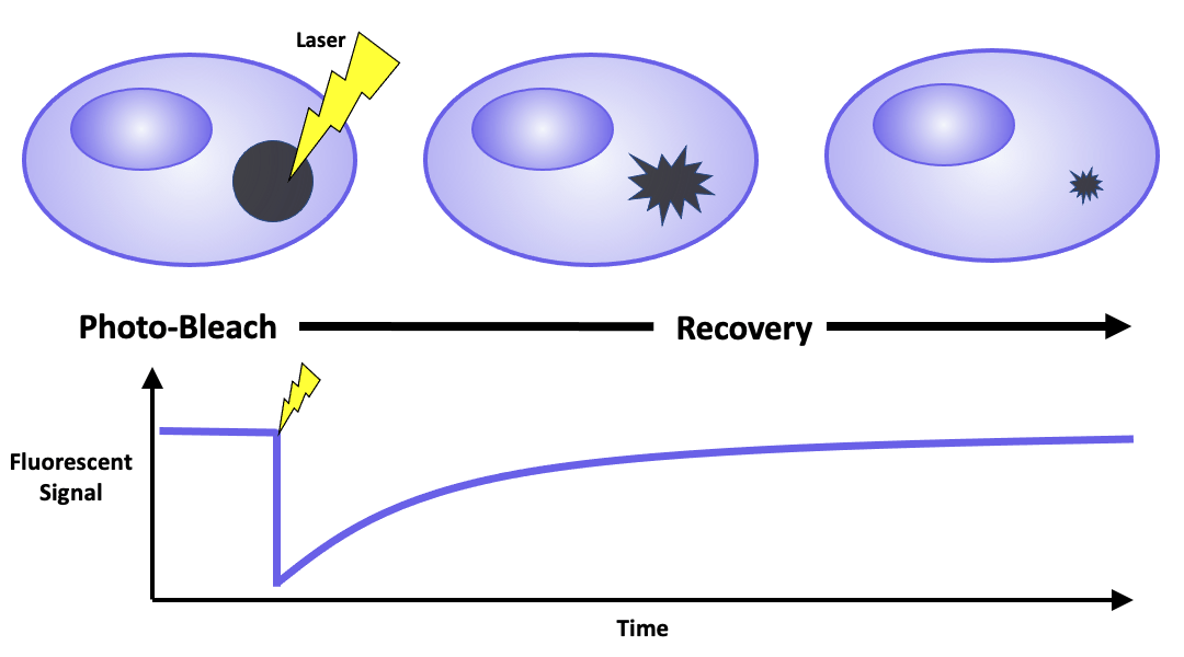
When a fluorophore absorbs light, it enters an excited state. While most fluorophores quickly return to their ground state by emitting light, some undergo side reactions that lead to the formation of reactive oxygen species (ROS). These ROS can then damage the fluorophore, causing it to lose its ability to fluoresce permanently.
According to a review in Nature Methods, the rate of photobleaching is directly proportional to the intensity of the excitation light and the duration of exposure. Furthermore, certain environmental factors, such as the presence of oxygen and the pH of the sample, can also influence the rate of photobleaching.
The Impact on Research: Challenges and Limitations
The limitations imposed by photobleaching are far-reaching. In live-cell imaging, where researchers aim to observe dynamic processes over time, photobleaching can obscure subtle changes in fluorescence intensity, making it difficult to accurately track cellular events.
Quantifying protein expression using fluorescence intensity becomes unreliable as the signal degrades over time. This can lead to inaccurate conclusions about the abundance and distribution of proteins within cells.
Dr. Emily Carter, a professor of cell biology at Stanford University, explains: "Photobleaching can introduce significant artifacts into our data, especially when we're trying to measure subtle changes in protein dynamics. It's crucial to implement strategies to minimize its impact."
Mitigating Photobleaching: Strategies and Techniques
Researchers have developed various strategies to minimize photobleaching and improve the quality and duration of fluorescence microscopy experiments. One common approach is to reduce the intensity of the excitation light.
While this reduces the rate of photobleaching, it also reduces the signal intensity. Therefore, a balance must be struck between minimizing photobleaching and maintaining a detectable signal.
Another strategy is to use antifade reagents. These are chemical compounds that scavenge ROS, preventing them from damaging the fluorophores. Examples include glycerol, DABCO, and propyl gallate.
Spectral unmixing can help to separate out the signal from the fluorophore from the background fluorescence caused by damaged fluorophores. This mathematical technique can compensate for the impact of photobleaching on the image.
Advanced microscopy techniques such as spinning disk confocal microscopy and light sheet microscopy can also reduce photobleaching. These techniques illuminate only a small portion of the sample at a time, minimizing the overall exposure to excitation light.
Future Directions: Novel Fluorophores and Advanced Algorithms
The development of new fluorophores that are more resistant to photobleaching is an active area of research. Researchers are exploring new chemical structures and modifications that can enhance the photostability of fluorophores.
Furthermore, advanced image processing algorithms are being developed to correct for the effects of photobleaching. These algorithms can analyze the rate of photobleaching and compensate for the signal loss over time.
Dr. David Miller, a researcher at the National Institutes of Health (NIH), states, "The future of fluorescence microscopy lies in the development of more robust fluorophores and sophisticated computational tools that can mitigate the effects of photobleaching."
Ultimately, tackling photobleaching requires a multi-faceted approach. By combining strategies to minimize excitation light, using antifade reagents, and employing advanced image processing techniques, researchers can mitigate the impact of photobleaching and obtain more accurate and reliable data from fluorescence microscopy experiments.
The quest to overcome photobleaching continues, promising to unlock even deeper insights into the complexities of cellular life.