Shows Brain Activity By Tracking Glucose Absorption

For decades, scientists have sought a more direct and nuanced window into the intricate workings of the human brain. Current neuroimaging techniques, while powerful, often rely on indirect measures of neuronal activity. A groundbreaking new approach promises to revolutionize our understanding of brain function by visualizing glucose absorption in real-time.
This innovative technology, detailed in a recent publication in Nature Neuroscience, directly tracks glucose uptake in the brain. This provides a more granular and immediate picture of neuronal activity than ever before. The implications are vast, potentially transforming the diagnosis and treatment of neurological disorders, enhancing our comprehension of cognitive processes, and even offering insights into consciousness itself.
The Sweet Spot: Glucose as a Window to Brain Activity
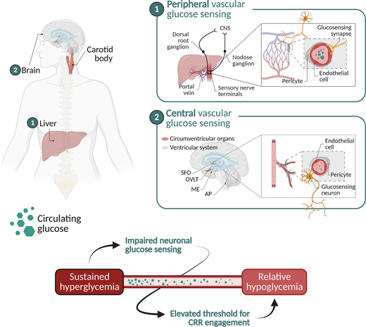
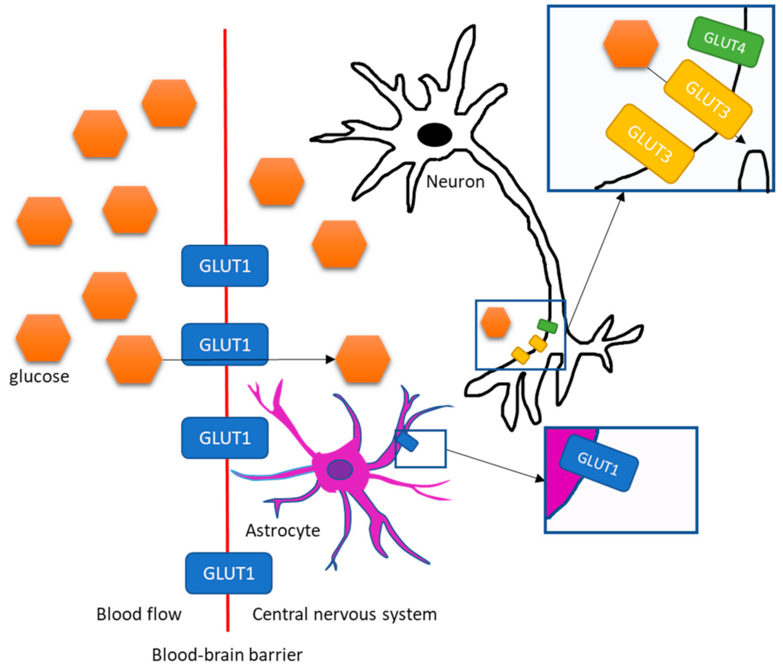
The brain, a notoriously energy-hungry organ, relies almost exclusively on glucose for fuel. When neurons fire, they consume glucose at an accelerated rate. This fundamental relationship is the cornerstone of this new neuroimaging technique.
Researchers have developed a specialized glucose analog that can be tracked using advanced imaging modalities. Unlike traditional methods that measure blood flow or oxygen consumption, this technique directly visualizes glucose metabolism at the cellular level. This offers unprecedented spatial and temporal resolution.
How It Works: A Deep Dive into the Technology
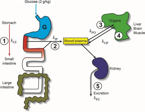
The process begins with the administration of a modified glucose molecule. This molecule is designed to be taken up by active neurons just like regular glucose. However, it contains a detectable tag, such as a fluorescent marker or a radioactive isotope.
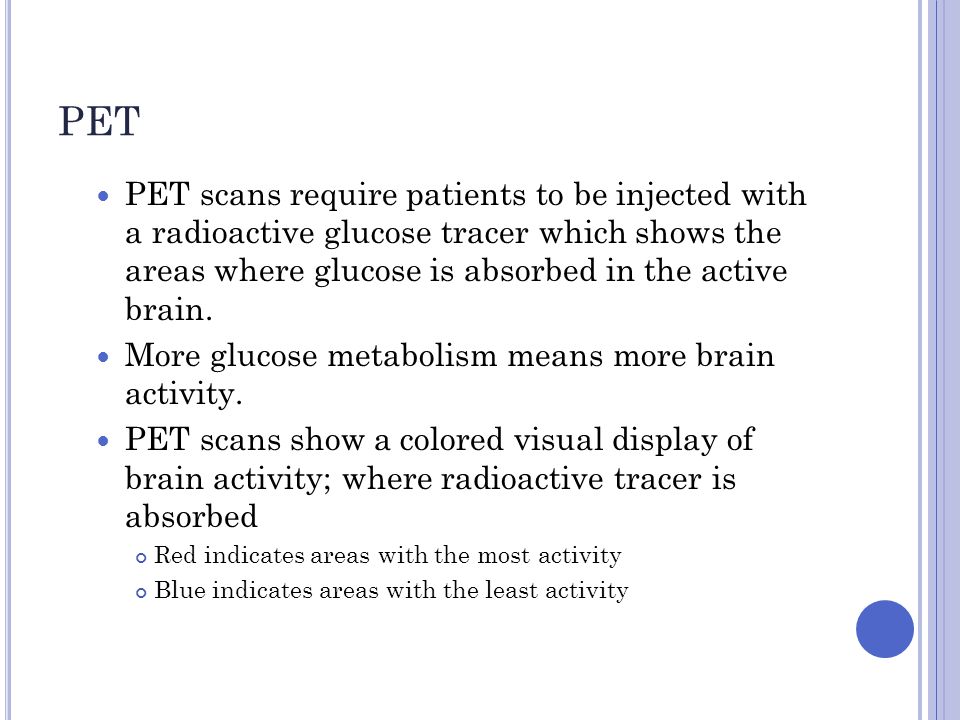
Once the analog is absorbed, advanced imaging technologies, like two-photon microscopy or positron emission tomography (PET), are used to map its distribution throughout the brain. The concentration of the tagged glucose analog directly corresponds to the level of neuronal activity in specific brain regions.
Dr. Anya Sharma, lead author of the Nature Neuroscience study, explained that this method offers several advantages over existing techniques. "fMRI, for example, relies on the blood-oxygen-level-dependent (BOLD) signal, which is an indirect measure of neuronal activity," she stated. "Our approach provides a more direct and immediate readout, allowing us to observe brain activity with greater precision."
Implications for Neurological Disorders
The ability to visualize glucose metabolism in real-time has profound implications for understanding and treating neurological disorders. Diseases like Alzheimer's, Parkinson's, and epilepsy are characterized by altered brain metabolism. Early detection of these metabolic changes could pave the way for more effective interventions.
For example, in Alzheimer's disease, glucose metabolism is known to decline in specific brain regions even before cognitive symptoms manifest. This new imaging technique could potentially detect these early metabolic deficits, allowing for earlier diagnosis and potentially slowing down disease progression.
Epilepsy research could also greatly benefit. Identifying areas of increased glucose metabolism during seizures with higher precision could help guide surgical interventions, leading to better outcomes for patients with drug-resistant epilepsy. Researchers are hopeful this will pinpoint seizure origin with higher accuracy.
Beyond Disease: Unlocking the Secrets of Cognition
Beyond neurological disorders, this new technology offers exciting possibilities for studying cognitive processes. Researchers can now investigate how different brain regions work together during learning, memory, and decision-making with unprecedented detail.
Imagine being able to visualize the specific brain circuits activated when a person solves a complex problem or recalls a cherished memory. This level of understanding could revolutionize our approach to education, cognitive training, and even artificial intelligence. This could give way for better learning environments that stimulate the appropriate brain activity.
Challenges and Future Directions
Despite its immense potential, this new technique also faces several challenges. The glucose analogs used must be safe and non-toxic. The imaging technologies required are expensive and not widely available. There are ethical issues to consider with more granular and accurate mind reading.
Furthermore, the data generated by this technique is complex and requires sophisticated analytical tools. Addressing these challenges will be crucial for translating this technology into widespread clinical and research applications.
However, the research community is optimistic about the future. Ongoing efforts are focused on developing more sensitive and selective glucose analogs. Scientists also are working to improve the accessibility and affordability of the required imaging technologies.
"We are just at the beginning of this exciting journey," says Dr. Sharma. "With further refinement and validation, this technology has the potential to transform our understanding of the brain and revolutionize the treatment of neurological disorders."
Ethical Considerations
The capacity to observe brain activity with such detail raises significant ethical considerations. The potential for misuse of this technology, such as in mind reading or manipulation, needs careful consideration. The community must discuss possible abuse of the new found insights.
Robust ethical guidelines and regulations are essential to ensure that this powerful tool is used responsibly and for the benefit of society. This is a powerful technology with a potential to be abused if not handled carefully. The potential benefit outweighs the potential harm, but we must stay vigilant.
The Road Ahead
The development of a technique to visualize brain activity through glucose absorption represents a major breakthrough in neuroscience. While challenges remain, the potential benefits for understanding and treating neurological disorders, as well as unraveling the mysteries of cognition, are immense. The future of brain research looks brighter than ever.
As researchers continue to refine and validate this technology, we can expect to see a wave of new discoveries about the human brain and its remarkable capabilities. This promises a new era in brain imaging. Ultimately, it will lead to more effective treatments for the millions of people affected by neurological disorders worldwide.