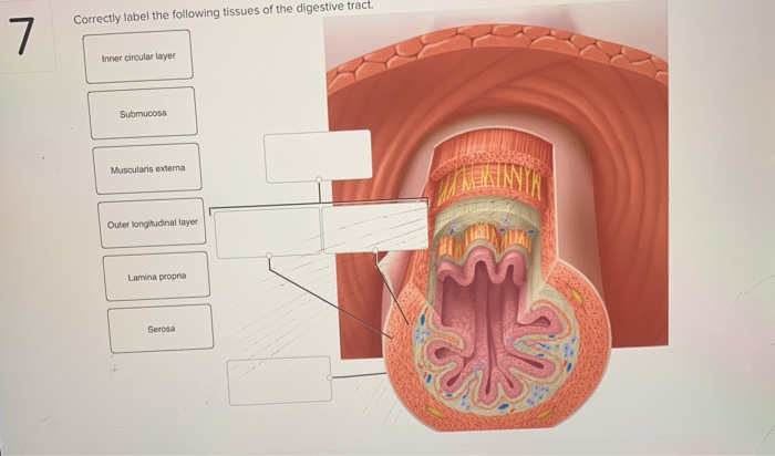
Correctly Label The Following Tissues Of The Digestive Tract.
Confusion and inaccuracies plague the understanding of digestive tract tissues, a foundational area of both biology and medicine. Misidentification of these tissues can lead to misdiagnosis, improper treatment, and a general undermining of patient care. The stakes are high, demanding a renewed focus on accurate anatomical knowledge.
This article delves into the critical importance of accurately identifying the digestive tract's tissue layers: the mucosa, submucosa, muscularis externa, and serosa/adventitia. We will explore the composition of each layer, common errors in identification, and the clinical consequences of these errors. Experts weigh in on best practices for education and the ongoing need for improved diagnostic accuracy.
The Building Blocks: Layers of the Digestive Tract
The Mucosa: The Inner Lining
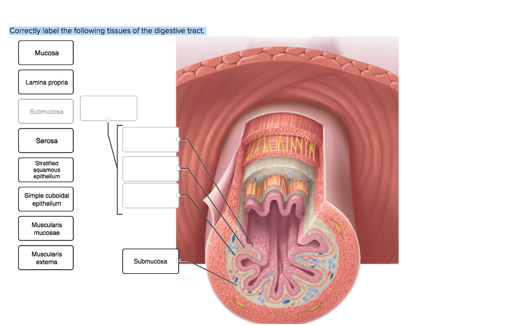
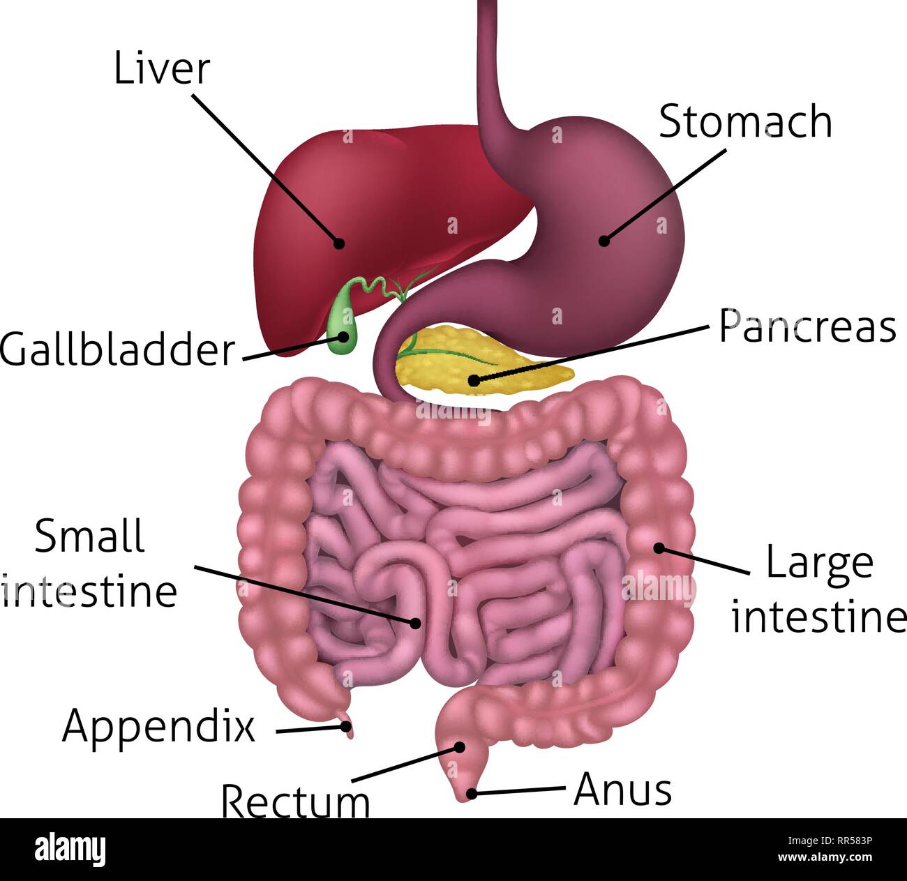
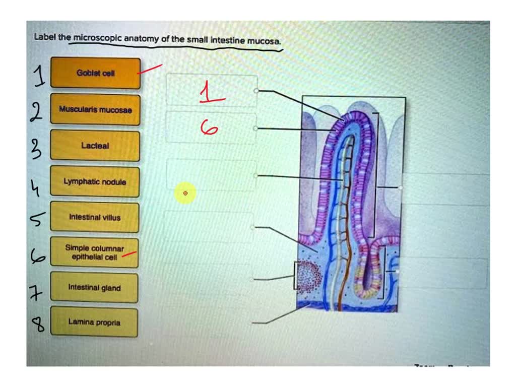
The mucosa is the innermost layer, in direct contact with the digested material. It’s composed of three sublayers: the epithelium, lamina propria, and muscularis mucosae. The epithelium is the absorptive and secretory layer, varying in structure along the tract.
The lamina propria is a layer of connective tissue containing blood vessels, lymphatics, and immune cells. Finally, the muscularis mucosae is a thin layer of smooth muscle that creates folds, increasing surface area.
The Submucosa: Support and Vasculature
The submucosa is a thicker layer of connective tissue beneath the mucosa. It contains larger blood vessels, lymphatic vessels, and nerves. This layer provides structural support and allows for distension.
In some regions, the submucosa also houses glands that secrete lubricating mucus. The submucosal plexus (Meissner's plexus), a network of nerve fibers, controls secretions and local blood flow.
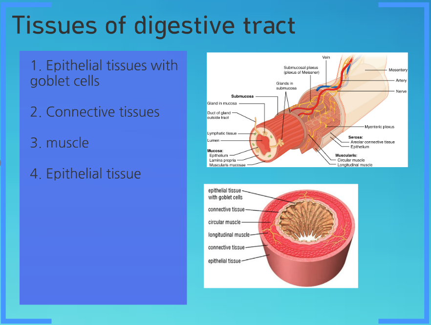
The Muscularis Externa: Motility
The muscularis externa is responsible for the digestive tract's motility. It typically consists of two layers of smooth muscle: an inner circular layer and an outer longitudinal layer. These layers contract and relax to propel food through the digestive system.
The myenteric plexus (Auerbach's plexus), located between these muscle layers, controls their contractions. Exceptions to this two-layer structure exist, such as the stomach, which has an additional oblique layer.
The Serosa/Adventitia: The Outer Layer
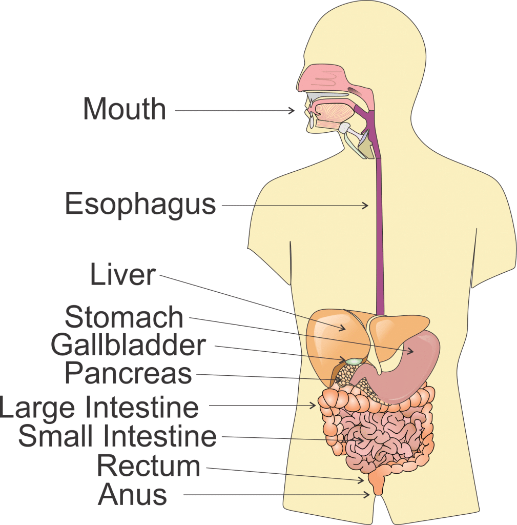
The outermost layer is either the serosa or the adventitia. The serosa is a serous membrane, present in regions suspended in the abdominal cavity. It provides a smooth surface and reduces friction.
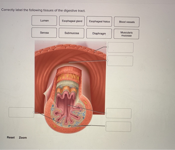
The adventitia is a layer of connective tissue that anchors the digestive tract to surrounding structures. It is found in regions that are not freely suspended, such as the esophagus.
Common Errors in Tissue Identification
One common mistake is confusing the muscularis mucosae of the mucosa with the muscularis externa. The muscularis mucosae is much thinner and is located within the mucosa itself.
Another error involves misidentifying the submucosa. Its characteristic presence of larger blood vessels and the submucosal plexus is often overlooked. This can lead to an incorrect assessment of tumor invasion depth.
Distinguishing between the serosa and adventitia can also be challenging. Understanding the location of the digestive tract segment is crucial. If it is within the peritoneal cavity, it should be serosa; if attached to other tissues, it is likely adventitia.
Clinical Consequences of Misidentification
Inaccurate tissue identification can have serious clinical implications. For example, misinterpreting the depth of tumor invasion in a biopsy sample can lead to incorrect staging of cancer. This can impact treatment decisions and prognosis.
If the muscular layers are misinterpreted, the diagnosis can be incorrect. An example is mistaking inflammatory bowel disease with other conditions.
Moreover, incorrect identification can lead to unnecessary surgeries or ineffective medical treatments. These consequences underscore the importance of accurate diagnosis.
Expert Perspectives on Improvement
Dr. Emily Carter, a professor of pathology at the University of California, San Francisco, emphasizes the need for thorough training. "Medical students and residents must be provided with ample opportunities to examine histological slides and learn from experienced pathologists," she states.
Dr. David Lee, a gastroenterologist at Massachusetts General Hospital, advocates for the use of advanced imaging techniques. "Techniques like confocal microscopy and optical coherence tomography can provide higher resolution images, aiding in tissue identification," he suggests.
Several organizations are working to standardize terminology and diagnostic criteria. They create guides to help prevent misidentification of these tissues.
Looking Ahead: Ensuring Accurate Diagnosis
Addressing the challenge of accurately identifying digestive tract tissues requires a multi-faceted approach. This includes enhanced training, the adoption of advanced diagnostic tools, and standardized reporting practices. Continuous education and collaboration among pathologists, gastroenterologists, and other healthcare professionals are essential.
Ultimately, improving diagnostic accuracy will lead to better patient outcomes. It will reduce unnecessary procedures and improve the effectiveness of treatment strategies.
By prioritizing accurate anatomical knowledge, we can ensure that patients receive the best possible care.