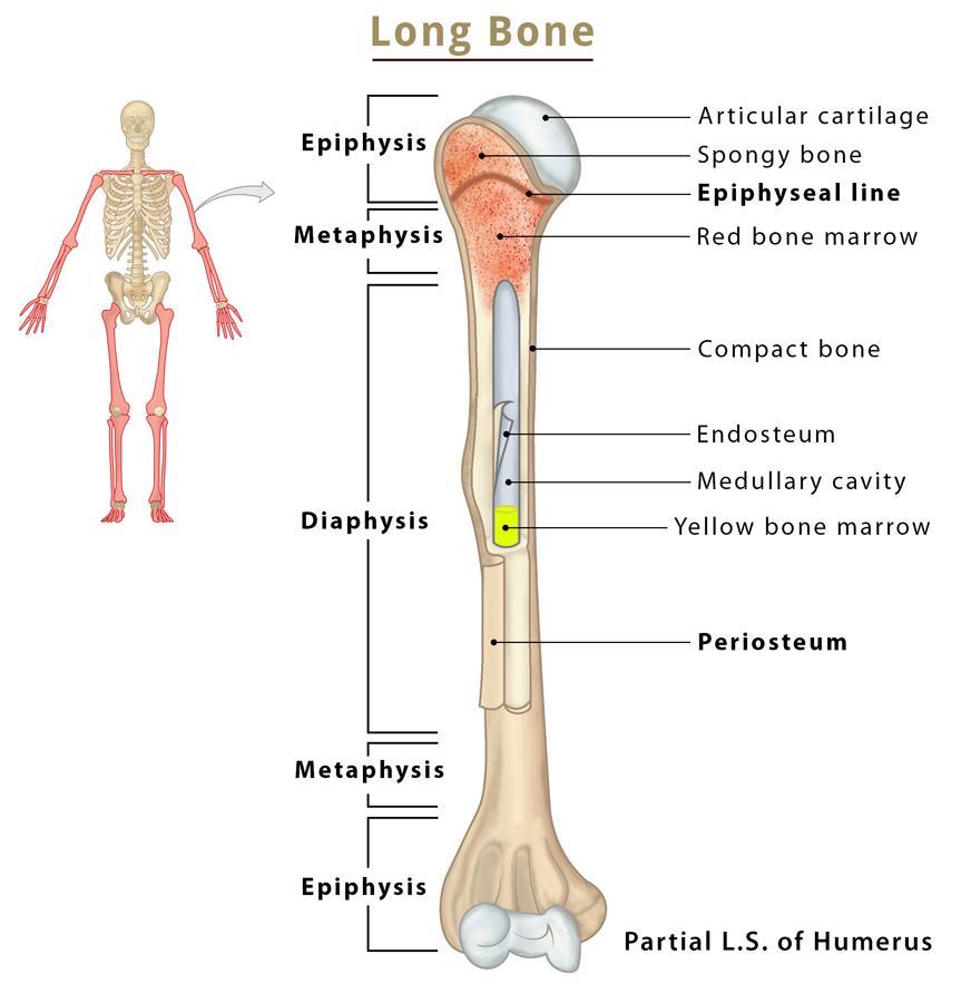
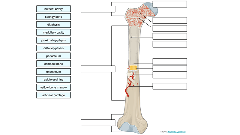
Label The Structures Of The Bone.
Imagine holding a smooth, sun-bleached bone in your hands. Its intricate curves and subtle textures whisper tales of resilience, strength, and the silent framework that supports life. But beyond its aesthetic appeal, this bone is a complex architectural marvel, a living scaffold brimming with secrets just waiting to be unveiled.
Understanding the anatomy of bones, labeling their diverse structures, isn't just a lesson in biology; it's a pathway to appreciating the incredible design and functionality of the human body. This article will guide you through the fascinating world of bone structure, highlighting key components and explaining their vital roles in maintaining our health and well-being. We'll explore everything from the dense outer layer to the spongy interior, revealing the dynamic processes that keep our skeletons strong and adaptable.
The Bone's Foundation: A Look Inside
Bones are far from being inert, lifeless objects. They are dynamic tissues, constantly being remodeled and rebuilt by specialized cells. This ongoing process allows bones to adapt to stress, heal from injuries, and contribute to the overall health of the body.
At the most basic level, bone is composed of a matrix of collagen fibers and mineral deposits, primarily calcium phosphate. This combination gives bone its remarkable strength and flexibility, allowing it to withstand significant forces without fracturing.
Periosteum: The Outer Guardian
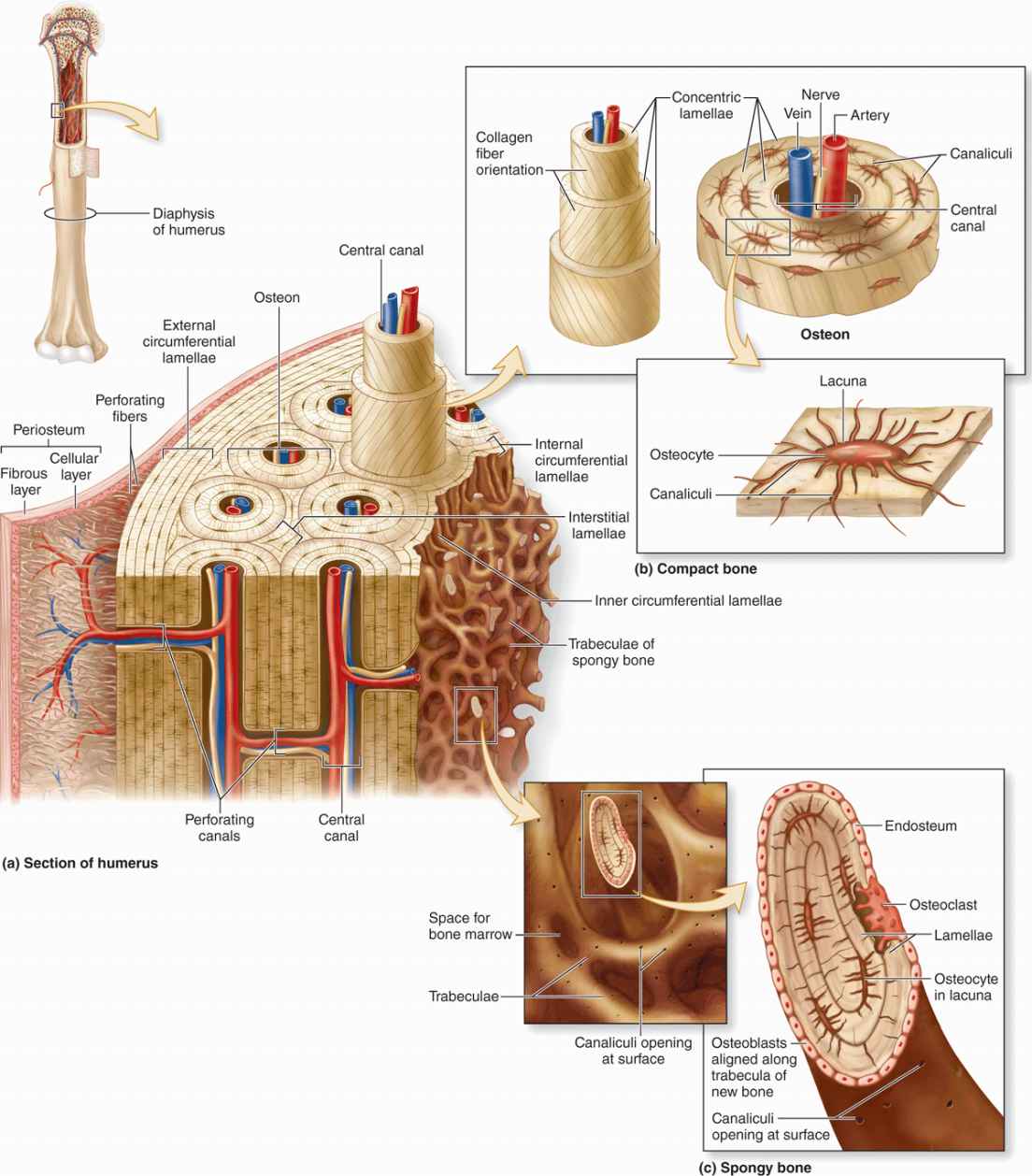
The periosteum is a tough, fibrous membrane that covers the outer surface of most bones. This protective layer is rich in blood vessels and nerves, providing essential nutrients and sensory information to the bone tissue.
It also plays a critical role in bone growth and repair, containing cells that can differentiate into bone-forming cells, known as osteoblasts. The periosteum is particularly thick in areas where tendons and ligaments attach to bone, providing a strong anchor point for these structures.
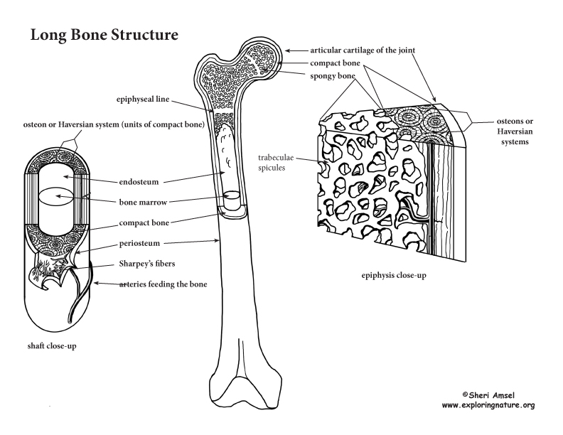
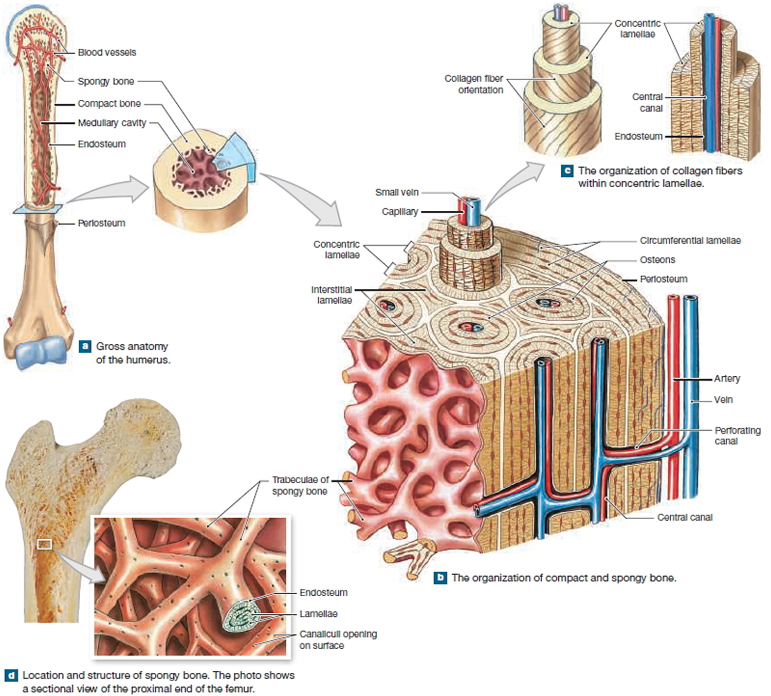
Compact Bone: Strength and Stability
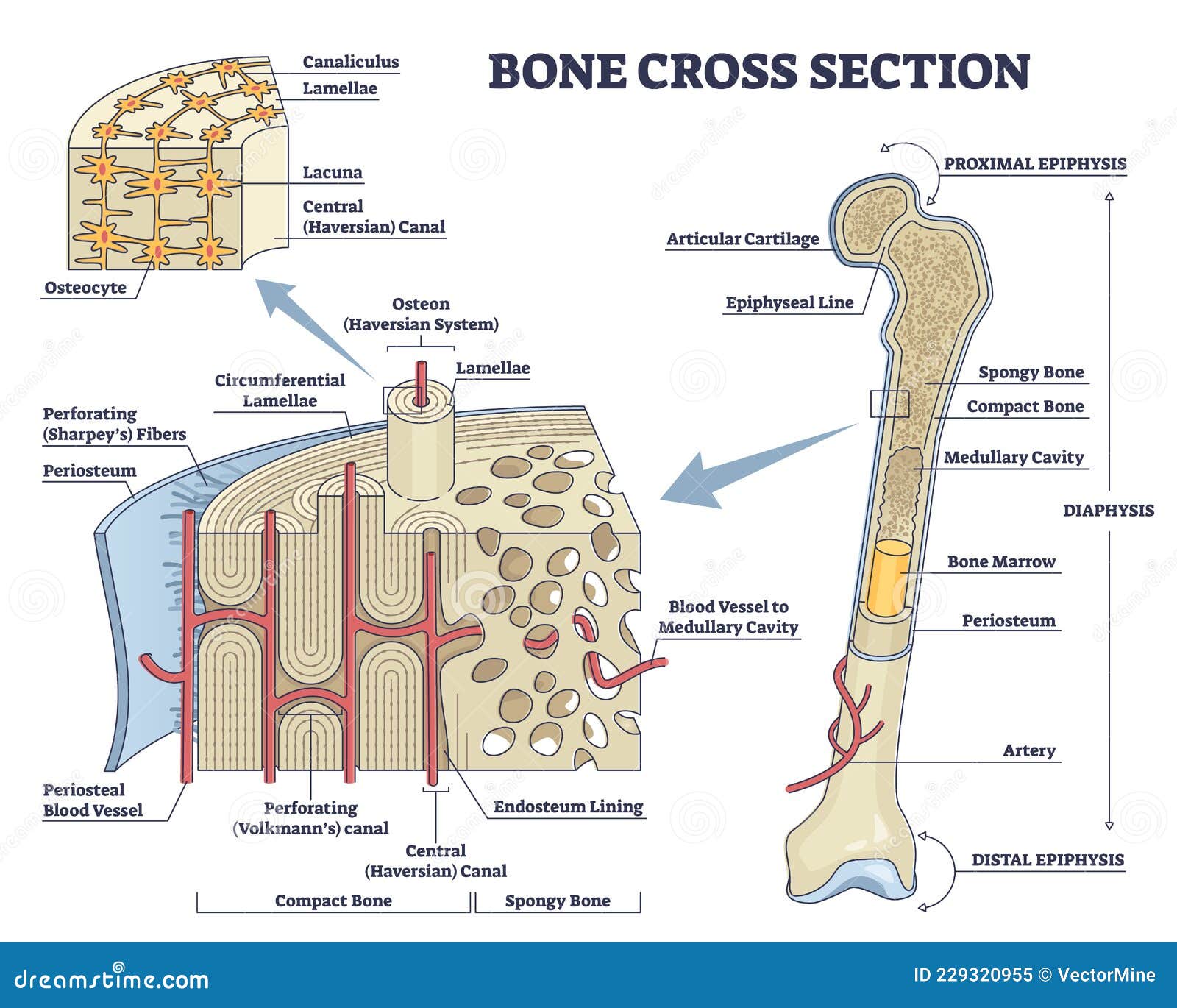
Beneath the periosteum lies the compact bone, also known as cortical bone. This dense, solid layer forms the outer shell of most bones, providing strength and resistance to bending and twisting forces.
Compact bone is organized into cylindrical structures called osteons or Haversian systems. Each osteon consists of concentric layers of bone tissue, called lamellae, surrounding a central canal that contains blood vessels and nerves. These canals, known as Haversian canals, allow nutrients and waste products to be transported throughout the bone.
These osteons are tightly packed together, giving compact bone its characteristic density and strength. Tiny channels called canaliculi connect the osteocytes, bone cells embedded within the lacunae of each lamella. This intricate network enables communication and nutrient exchange between bone cells.
Spongy Bone: Lightweight Support
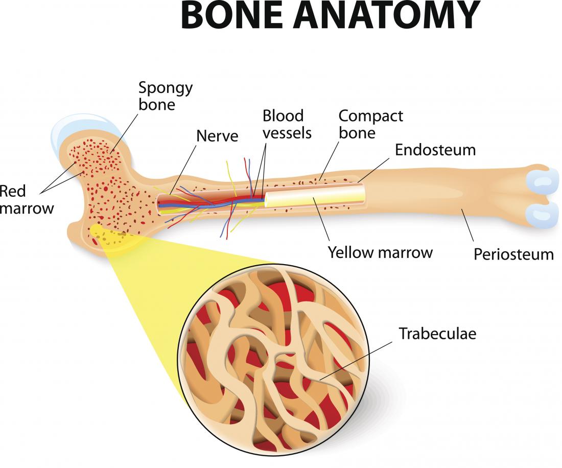
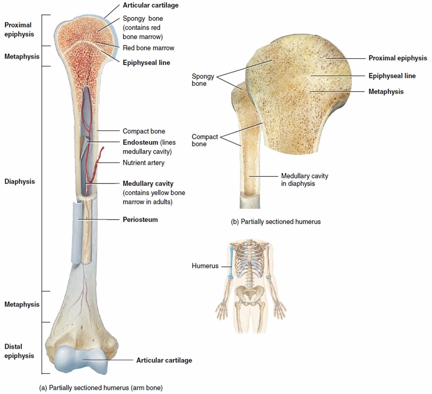
Inside the compact bone, particularly at the ends of long bones and within the vertebrae, is the spongy bone, also known as cancellous bone. This type of bone has a porous, honeycomb-like structure, which makes it lighter than compact bone while still providing significant strength and support.
The spaces within spongy bone are filled with bone marrow, the site of blood cell formation. Spongy bone is arranged in a network of bony struts called trabeculae. The trabeculae are aligned along lines of stress, providing maximum strength with minimal weight.
The trabecular network also helps to distribute forces evenly throughout the bone, preventing stress concentrations that could lead to fractures. This design is a remarkable example of structural optimization in nature.
Bone Features: Landmarks and Function
Bones exhibit a variety of surface features, or bone markings, that serve different purposes. These features include projections, depressions, and openings, each adapted to specific functions such as muscle attachment, joint articulation, or passage of blood vessels and nerves.
Processes are bony projections that serve as attachment sites for muscles, tendons, and ligaments. Examples include the trochanters of the femur (thigh bone) and the spinous processes of the vertebrae.
Depressions are indentations or hollow areas in the bone surface. These features often accommodate other structures, such as blood vessels or nerves. Examples include the fossa, sulcus, and foramen.
Foramina are openings or holes in the bone that allow blood vessels and nerves to pass through. The foramen magnum in the skull, for example, allows the spinal cord to connect to the brain. The nutrient foramen allows blood vessels to enter the bone.
Articular surfaces are smooth, cartilage-covered areas where bones articulate with each other to form joints. These surfaces are typically located at the ends of long bones and on the facets of vertebrae.
Bone Cells: The Architects of the Skeleton
Bone tissue is composed of four main types of cells: osteoblasts, osteocytes, osteoclasts, and bone lining cells.
Osteoblasts are bone-forming cells that synthesize and secrete the organic matrix of bone, called osteoid. They also play a role in the mineralization of bone tissue.
Once osteoblasts become surrounded by bone matrix, they differentiate into osteocytes. These cells are embedded within small cavities called lacunae and are responsible for maintaining the bone matrix. They communicate with each other through canaliculi.
Osteoclasts are large, multinucleated cells that break down bone tissue through a process called bone resorption. This process is essential for bone remodeling and repair.
Bone lining cells are found on the surface of bones and are thought to regulate the movement of calcium and phosphate into and out of the bone. These cells are derived from osteoblasts.
The Dynamic Skeleton: A Living Framework
Bones are not static structures; they are constantly being remodeled and rebuilt throughout life. This dynamic process, called bone remodeling, involves the coordinated activity of osteoblasts and osteoclasts.
Bone remodeling allows bones to adapt to changing mechanical stresses, repair injuries, and maintain calcium homeostasis. Wolff's Law states that bone adapts to the loads it is placed under. Bone deposition happens where needed and bone resorption occurs where there is little stress. Bone remodeling is influenced by a variety of factors, including hormones, vitamins, and physical activity.
Understanding the structure and function of bones is crucial for maintaining skeletal health throughout life. A balanced diet, regular exercise, and adequate vitamin D intake are essential for promoting strong, healthy bones.
A Final Reflection
Exploring the intricate world of bone structure reveals the remarkable complexity and resilience of the human body. From the tough outer periosteum to the spongy inner trabeculae, each component plays a vital role in supporting our movements, protecting our organs, and maintaining our overall health. By learning to label and appreciate these structures, we gain a deeper understanding of ourselves and the incredible architecture that allows us to thrive.
Next time you consider your skeleton, remember the tireless work of osteoblasts, osteocytes, and osteoclasts, constantly remodeling and rebuilding the framework that supports you. Appreciate the elegant design of the trabecular network, the strength of the compact bone, and the protective embrace of the periosteum. The skeleton is more than just bones; it is a living testament to the power and beauty of biological engineering.
![Label The Structures Of The Bone. [DIAGRAM] Diagram Of Bone With Labels - MYDIAGRAM.ONLINE](http://image.shutterstock.com/z/stock-vector-illustration-of-anatomy-of-bone-with-label-on-abstract-background-78759112.jpg)