Magnifying Power Of Compound Microscope Class 12

For generations of students, the compound microscope has been the quintessential tool for exploring the unseen world. It holds a prominent place in biology education, particularly at the Class 12 level, where understanding its magnifying power is crucial for grasping cellular structures and microscopic organisms.
This article delves into the intricacies of calculating the magnifying power of a compound microscope, a fundamental concept for students and educators alike. The microscope's ability to enlarge images allows students to observe minute details of biological specimens.
Understanding Magnification: Key Concepts
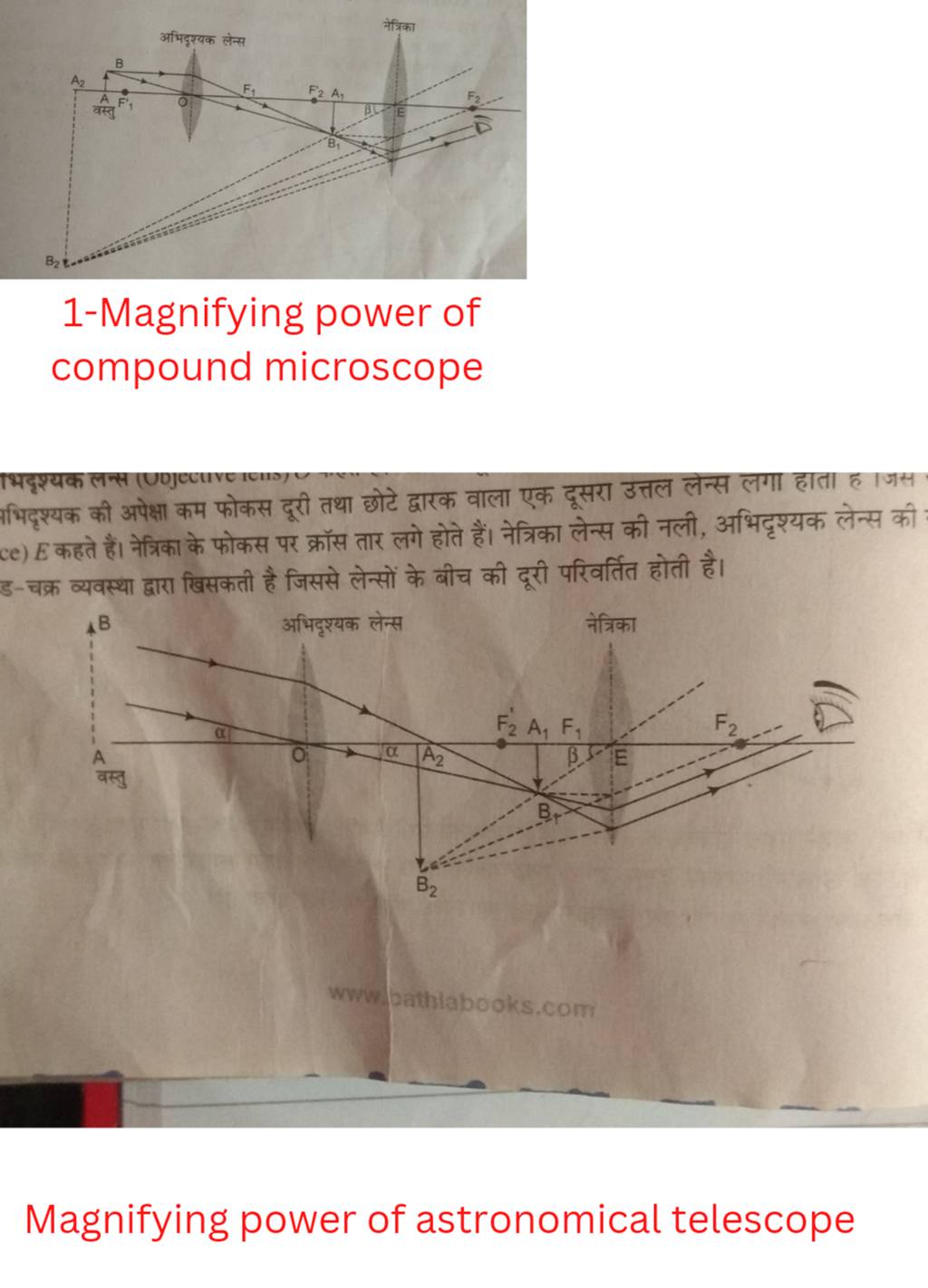
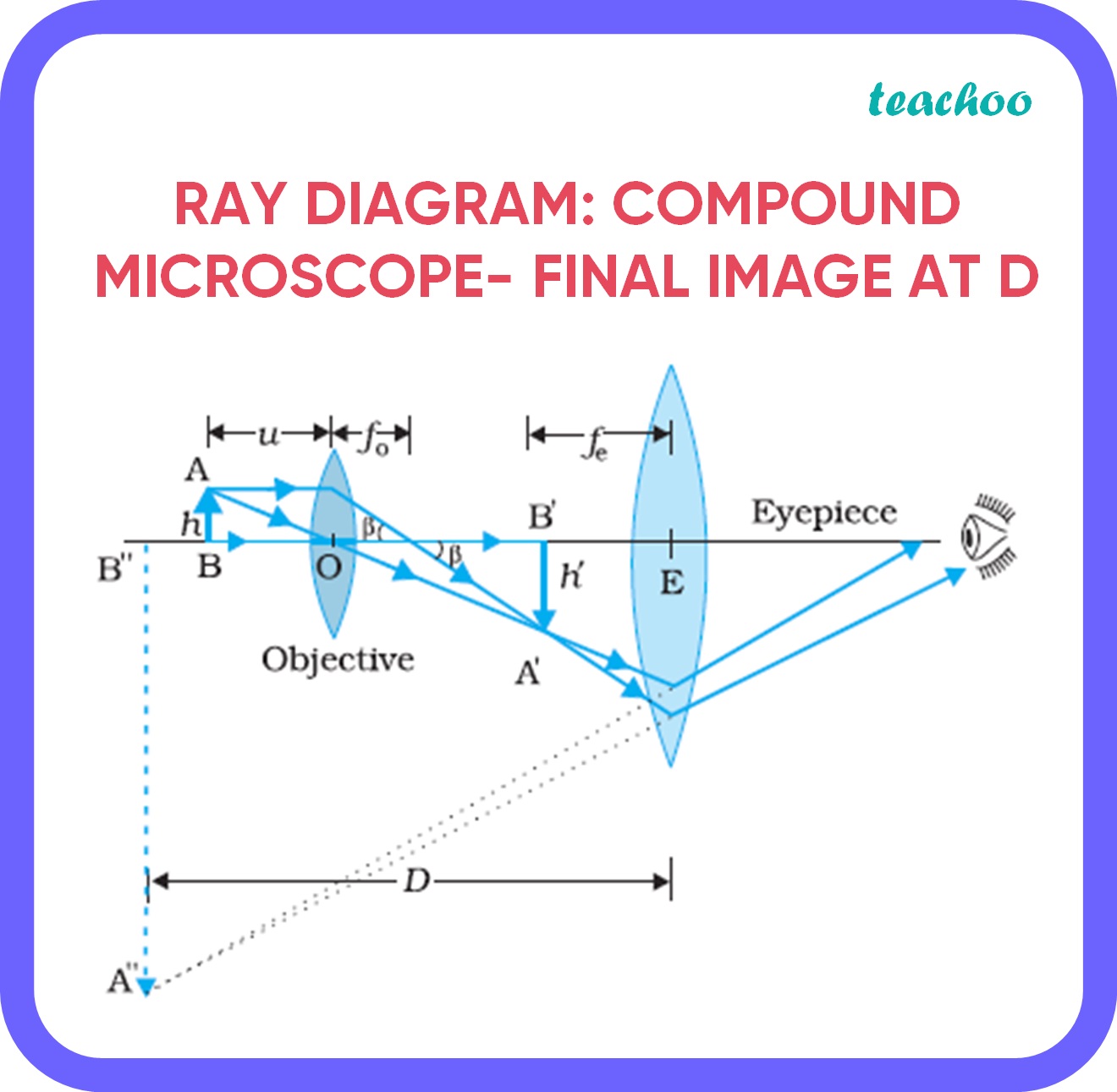
The magnifying power of a compound microscope is not simply a single number. It is a result of the combined effect of two lenses: the objective lens and the eyepiece lens.
The objective lens, positioned closest to the specimen, produces an initial magnified image. This image is then further magnified by the eyepiece lens, also known as the ocular lens, which the observer looks through.
Calculating Total Magnification
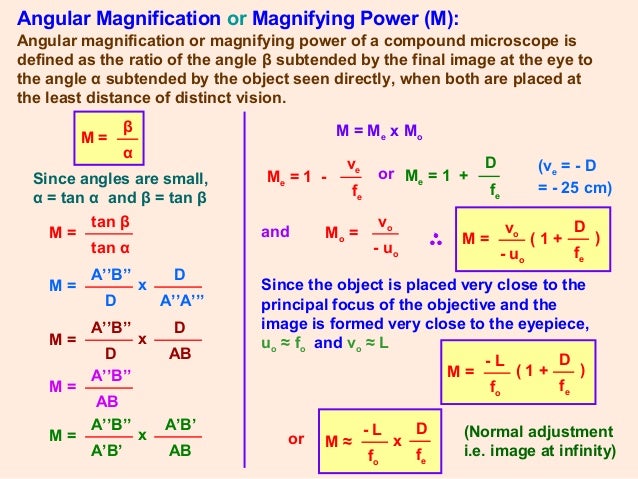
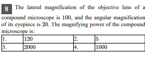
To determine the total magnification of a compound microscope, one multiplies the magnification of the objective lens by the magnification of the eyepiece lens. Mathematically, this is expressed as:
Total Magnification = (Magnification of Objective Lens) × (Magnification of Eyepiece Lens).
For instance, if an objective lens has a magnification of 40x and the eyepiece lens has a magnification of 10x, the total magnification is 400x. This means the image observed appears 400 times larger than its actual size.
Understanding this calculation is a core competency for Class 12 biology students, enabling them to accurately interpret microscopic observations. It also allows them to compare the effectiveness of different lens combinations.
Practical Applications in Biology Education
The concept of magnification is not merely theoretical; it has profound practical implications in biology education. Students use microscopes to observe cells, tissues, and microorganisms, furthering their understanding of life at the microscopic level.
In practical laboratory exercises, students often prepare slides of onion peel cells or cheek cells and observe them under varying magnifications. These activities solidify their understanding of cellular structures like the nucleus, cytoplasm, and cell wall.
By manipulating the objective lenses and eyepieces, students can observe the same specimen at different magnifications. This helps them appreciate the level of detail discernible at each magnification.
Challenges and Considerations
While calculating magnification is straightforward, there are nuances to consider for optimal microscopic observation. Simply increasing magnification does not always improve the quality of the image.
Beyond a certain point, higher magnification can result in a blurred or distorted image due to limitations in the microscope's resolution. Resolution refers to the microscope's ability to distinguish between two closely spaced objects.
Illumination is also a critical factor. Proper lighting is essential for achieving clear and well-defined images, especially at higher magnifications.
The Impact on Scientific Understanding
The compound microscope, and the understanding of its magnifying power, has revolutionized biology and medicine. It has allowed scientists to visualize microorganisms, diagnose diseases, and develop new treatments.
The foundation laid in Class 12 biology provides students with the necessary skills and knowledge to pursue careers in these fields. Many go on to become researchers, doctors, and other healthcare professionals.
By mastering the principles of microscopy, students are empowered to explore the microscopic world and contribute to scientific advancements.