Which Of The Following Substances Regulates Muscle Actions
The human body, a marvel of biological engineering, orchestrates countless movements every moment, from the subtle blink of an eye to the powerful stride of a marathon runner. The question of precisely which substance governs these intricate muscle actions sparks ongoing research and reveals a complex interplay of physiological processes. Getting to the heart of this issue requires a deep dive into the world of neurotransmitters, ions, and energy molecules that work in concert to enable every twitch, flex, and contraction.
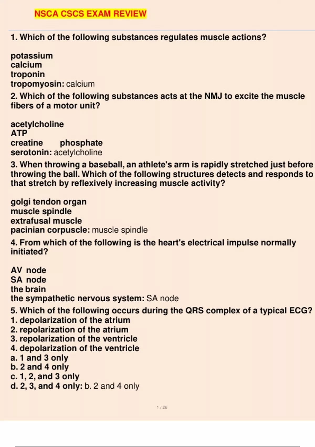
At its core, muscle action regulation isn't a solo performance by a single substance. Instead, it is a carefully choreographed process where multiple key players each play a crucial role. This article will explore the roles of calcium ions, acetylcholine, ATP (adenosine triphosphate), and other vital components in the symphony of muscle control.
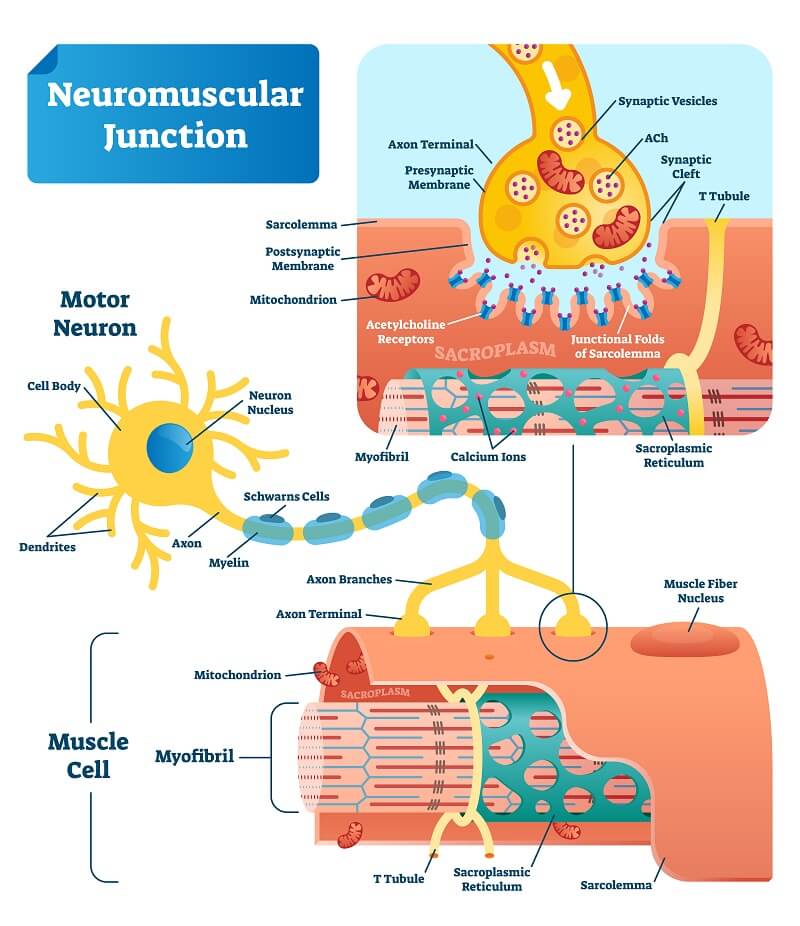
The Prime Mover: Acetylcholine and Neuromuscular Transmission
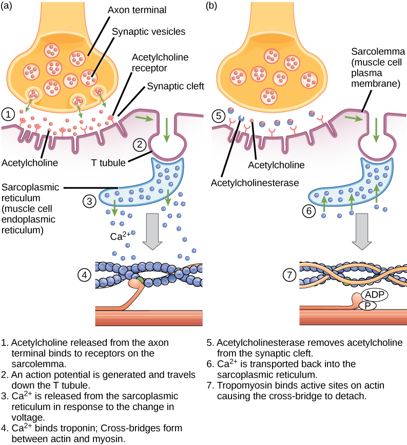
The story of muscle action begins at the neuromuscular junction, the meeting point between a motor neuron and a muscle fiber. Here, acetylcholine, a neurotransmitter, acts as the primary messenger.
When a nerve impulse arrives, it triggers the release of acetylcholine into the synaptic cleft, the small gap separating the neuron and muscle fiber.
Acetylcholine then binds to receptors on the muscle fiber membrane, initiating a chain of events that ultimately leads to muscle contraction.
Depolarization and Action Potentials
The binding of acetylcholine opens ion channels, allowing sodium ions to flow into the muscle fiber. This influx of positive charge causes depolarization, a change in the electrical potential across the membrane.
If the depolarization reaches a threshold, it triggers an action potential, an electrical signal that travels along the muscle fiber.
This action potential is crucial for propagating the signal deep within the muscle cell, ensuring a coordinated contraction.
Calcium: The Trigger for Contraction
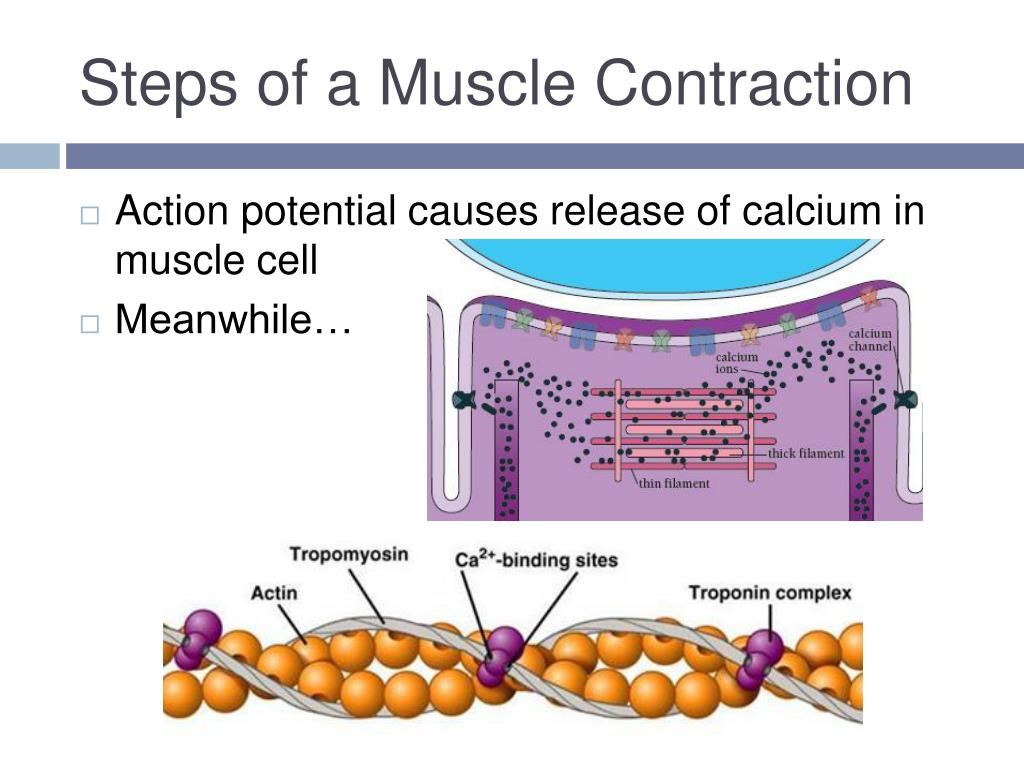
The action potential traveling along the muscle fiber stimulates the release of calcium ions (Ca2+) from the sarcoplasmic reticulum, an intracellular storage site.
Calcium ions are the linchpin in the actual contractile process.
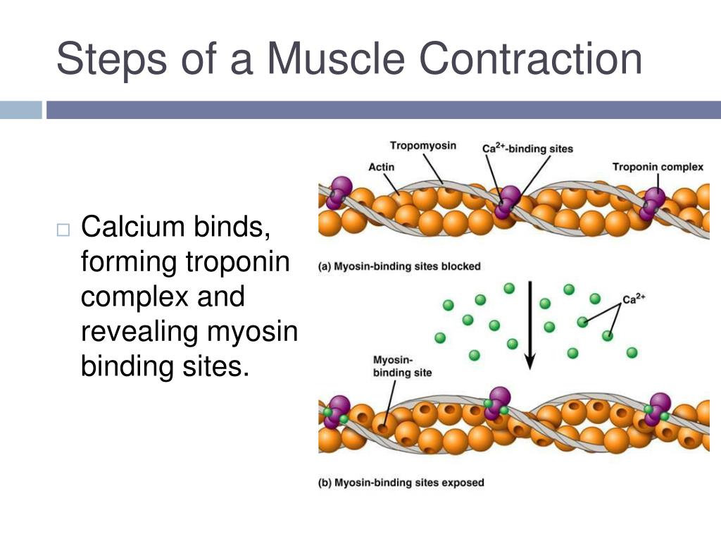
These ions bind to troponin, a protein complex located on actin filaments, the thin filaments within muscle fibers.
The Sliding Filament Mechanism
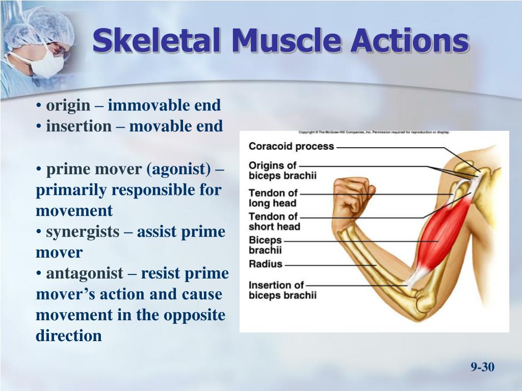
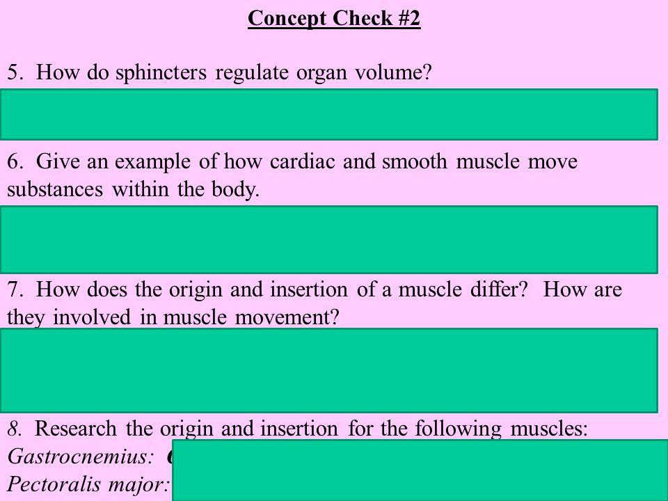
The binding of calcium to troponin causes a conformational change that exposes binding sites on actin. Myosin, the thick filament, can now bind to actin, forming cross-bridges.
Using energy from ATP, the myosin heads pull the actin filaments towards the center of the sarcomere, the basic contractile unit of the muscle fiber.
This sliding of filaments shortens the sarcomere, leading to muscle contraction.
ATP: The Energy Currency of Muscle Contraction
Adenosine triphosphate (ATP) is the primary energy source for muscle contraction.
ATP is required for several key steps, including the detachment of myosin from actin, the pumping of calcium back into the sarcoplasmic reticulum, and the maintenance of ion gradients across the muscle fiber membrane.
Without sufficient ATP, muscles cannot relax, leading to a state of rigor.
Creatine Phosphate: A Backup Energy System
The body uses creatine phosphate as a rapid source of ATP. During intense activity, creatine phosphate can quickly transfer a phosphate group to ADP (adenosine diphosphate) to regenerate ATP.
This system provides a short-term burst of energy before other metabolic pathways can ramp up.
However, creatine phosphate stores are limited and are quickly depleted during maximal effort.
Other Contributing Factors
While acetylcholine, calcium, and ATP are the central players, other substances also contribute to muscle action regulation.
For instance, electrolytes like sodium and potassium are essential for maintaining the proper electrical gradients across muscle fiber membranes.
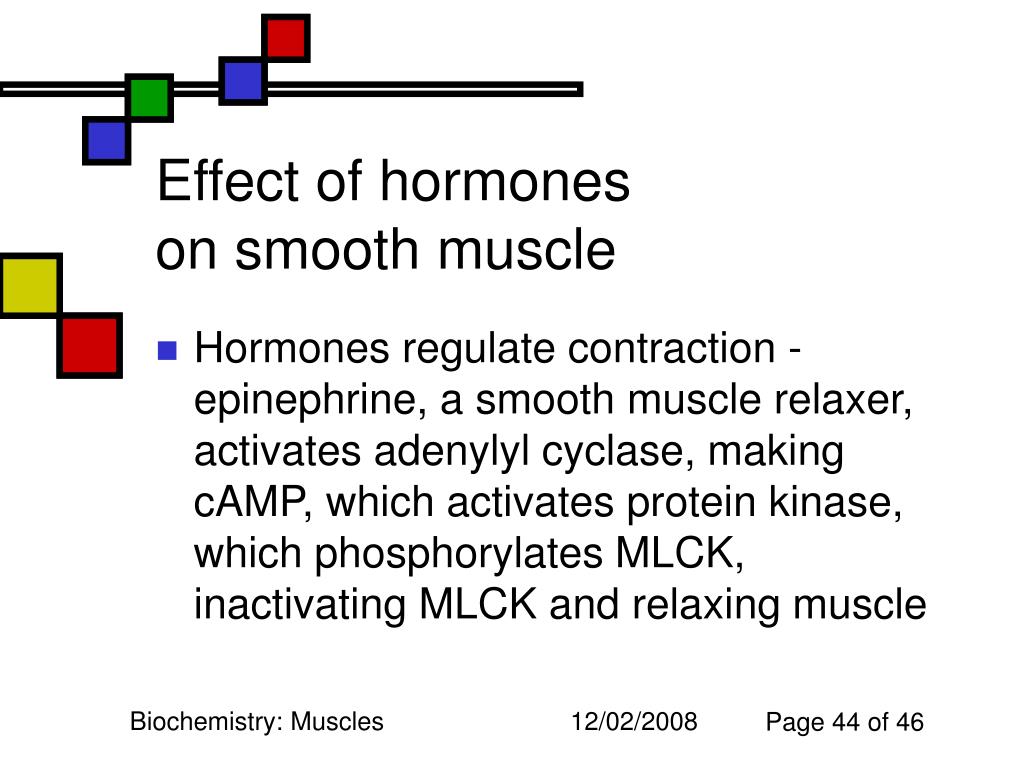
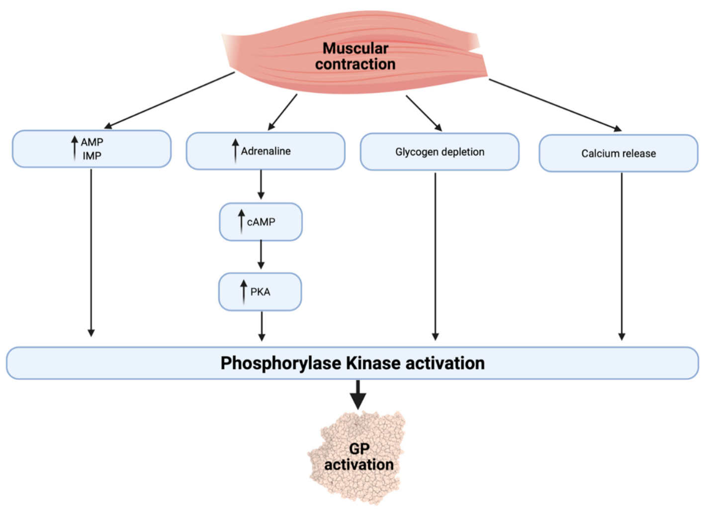
Hormones such as epinephrine and norepinephrine can influence muscle contractility and metabolism.
The Role of the Nervous System
The nervous system plays a critical role in coordinating muscle actions. Motor neurons transmit signals from the brain and spinal cord to muscles.
The frequency and pattern of these signals determine the strength and duration of muscle contractions.
Sensory feedback from muscles and joints also provides information to the nervous system, allowing for fine-tuning of movements.
Dysfunction and Disease
Disruptions in any of these substances or processes can lead to muscle dysfunction and disease. For example, myasthenia gravis is an autoimmune disorder where antibodies block acetylcholine receptors at the neuromuscular junction, leading to muscle weakness.
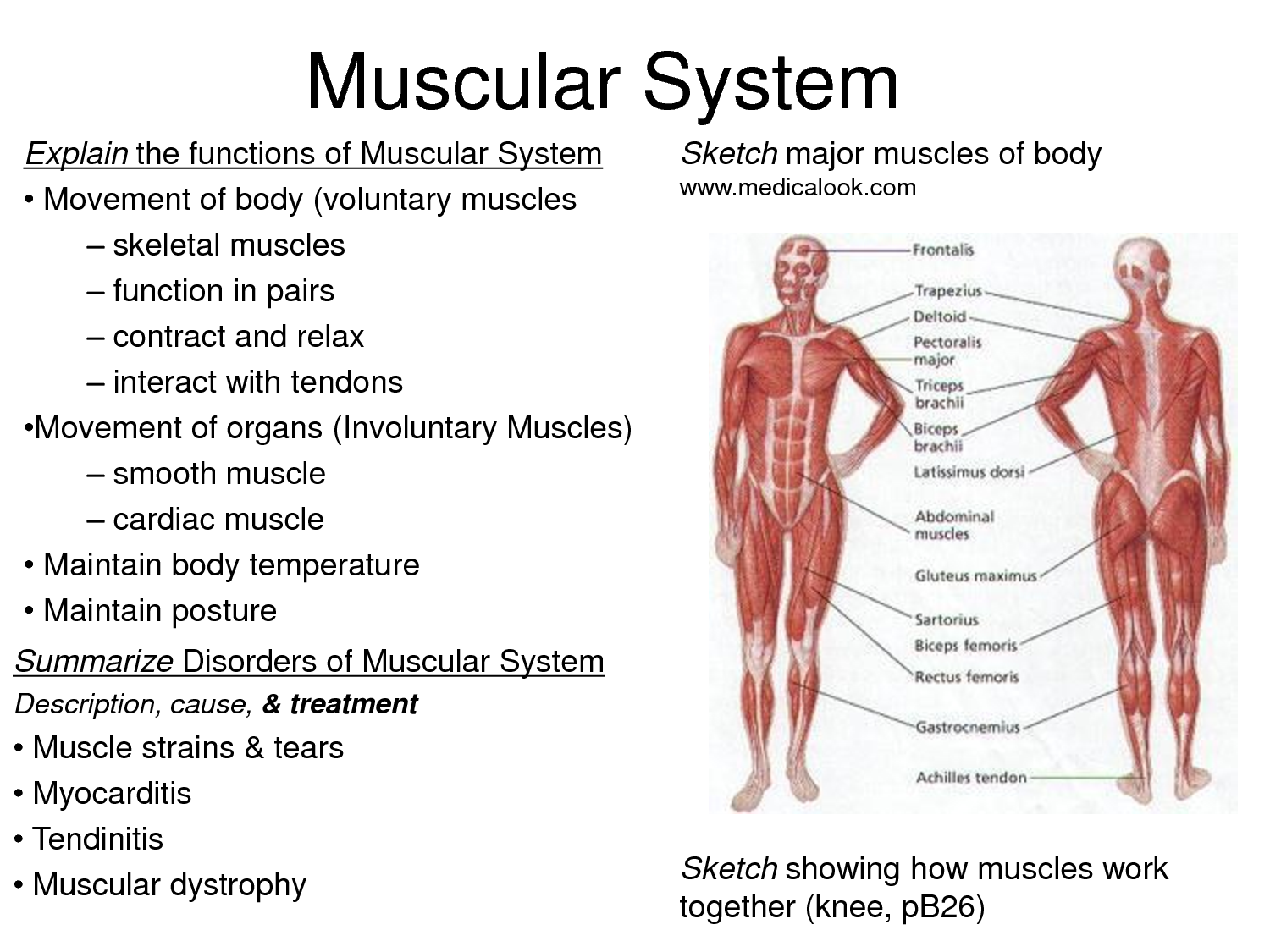
Muscular dystrophies are a group of genetic disorders that cause progressive muscle degeneration.
Electrolyte imbalances can also impair muscle function, leading to cramps or weakness.
The Future of Muscle Research
Research continues to explore the intricacies of muscle action regulation. Scientists are investigating new treatments for muscle diseases, developing strategies to enhance athletic performance, and exploring the potential of muscle tissue engineering.
Understanding the complex interplay of substances involved in muscle action is crucial for developing effective therapies for a wide range of conditions.
Further research promises to unlock even more secrets of the human body's remarkable ability to move.
In conclusion, while the question "Which substance regulates muscle actions?" might seem to point to a single answer, the reality is far more nuanced. Acetylcholine initiates the process, calcium triggers the contraction, and ATP fuels the entire operation. These substances, along with a host of other ions, electrolytes, and hormones, work in a coordinated fashion to control the incredible diversity of human movement. Further research promises even deeper insights into this complex and vital physiological process.